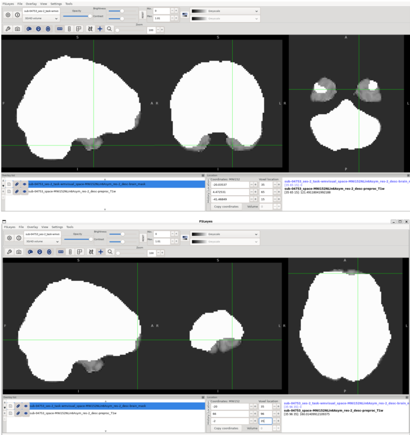
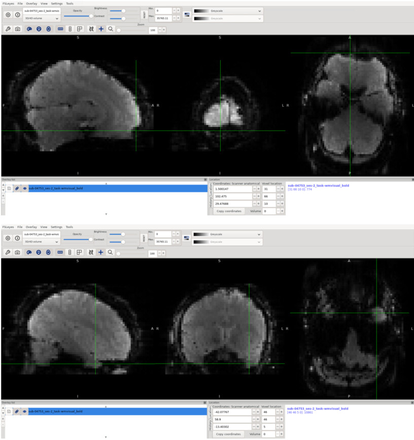
Hello, I’m new to fMRI preprocessing using fMRIPrep, and I have a question about the frontal and temporal part of the brain, which appears to be partially cut off in the preprocessed BOLD image.
I have posted both a T1 anatomical and T2 Bold scan for this subject. I have used a Siemens Prisma 3T scanner.
My questions are:
Is this expected behavior during BOLD preprocessing?
If this is a preprocessing issue, is there a way to improve or correct it?
Parameters of the T1 anatomical:
TR = 1.9 s, TE = 2.19 ms, TI = 0.9 s, flip angle = 9°, resolution/slice thickness = 1 mm, GRAPPA = 2
Parameters of the T2 bold:
TR = 1 s, TE = 30 ms, 3 mm slices, multiband factor = 3
Are those regions present in the original image? Also, at least for the frontal part, it could be a susceptibility distortion artifact, which can corrected if you have a fieldmap (or use something SynBOLD-DisCo for fieldmap-less correction).
Overlay the unmasked BOLD data. You will generally see signal dropout in the frontal and temporal poles, which is a susceptibility artifact distinct from the spatial distortion. Distortion correction as part of preprocessing can improve the localization of signal, but cannot restore attenuated signals.