Dear experts
I preprocessed task-based multi-echo fMRI data using fMRIPrep, and combined the echoes using tedana (ME-ICA). After registering to MNI standard space, I applied smoothing in SPM. However, when performing first-level analysis in SPM, the estimation failed. What could be the reasons for this?
06-Nov-2023 01:39:01 - Failed ‘Model estimation’
failed: spm_est_non_sphericity (line 208)
Please check your data: There are no significant voxels.
However, when I use the simple multi-echo fitting provided by fMRIPrep itself, there is no problem.
Hard to be sure - what does the SPM produced mask look like, for the model that failed. I think SPM makes that early in the process and you could see if it overlaps with your data appropriately/covers the brain after normalization.
Does the smoothed input into SPM look like a brain? As in, grayscale values with some range? Does it seem like it is reasonable data?
It looks like you are also using the denoised data. Did you inspect the output, including figures? The fmriprep data wouldn’t have been denoised (I don’t think…) and it is always possible that denoising removed your effects of interest. Given your task design it should be obvious if that is the case, as you would be able to see the block design.
I don’t think just turning off temporal auto-correlation is a solution.
In general my thought is that tedana inadvertently removed task components, so when you try and fit the model, there is nothing to fit - the task related BOLD is gone, but everything much be checked to be sure of things. For that, I think you should get much closer to your data to understand this problem, as in:
What does the task activation look like in the typical framework (fmriprep combined)? As in, the timecourses for voxels that are activated and what is the activation map like?
What does it look like when you turn off the AR(1) model with the denoised data? What is the map of activity - is it like the one above or missing things?
What components were removed by tedana? - there are figures for every identified component.
Do any of those components that were removed look like your task design? which seems like it should be fairly obvious.
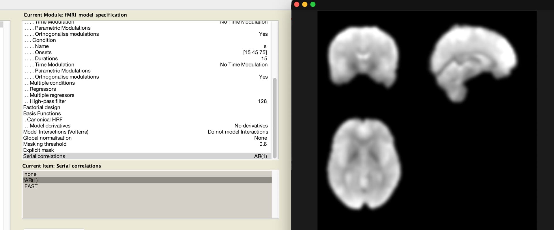
What does the SPM produced mask.nii file look like - does it match the whole brain you are showing in this image (this is related to the 0.8 “Masking threshold” you can see in the batch window". I’ve seen cases where SPM fails because the mask was too aggressive and there was nearly no data left to analyze.
Are you using the latest version of tedana?
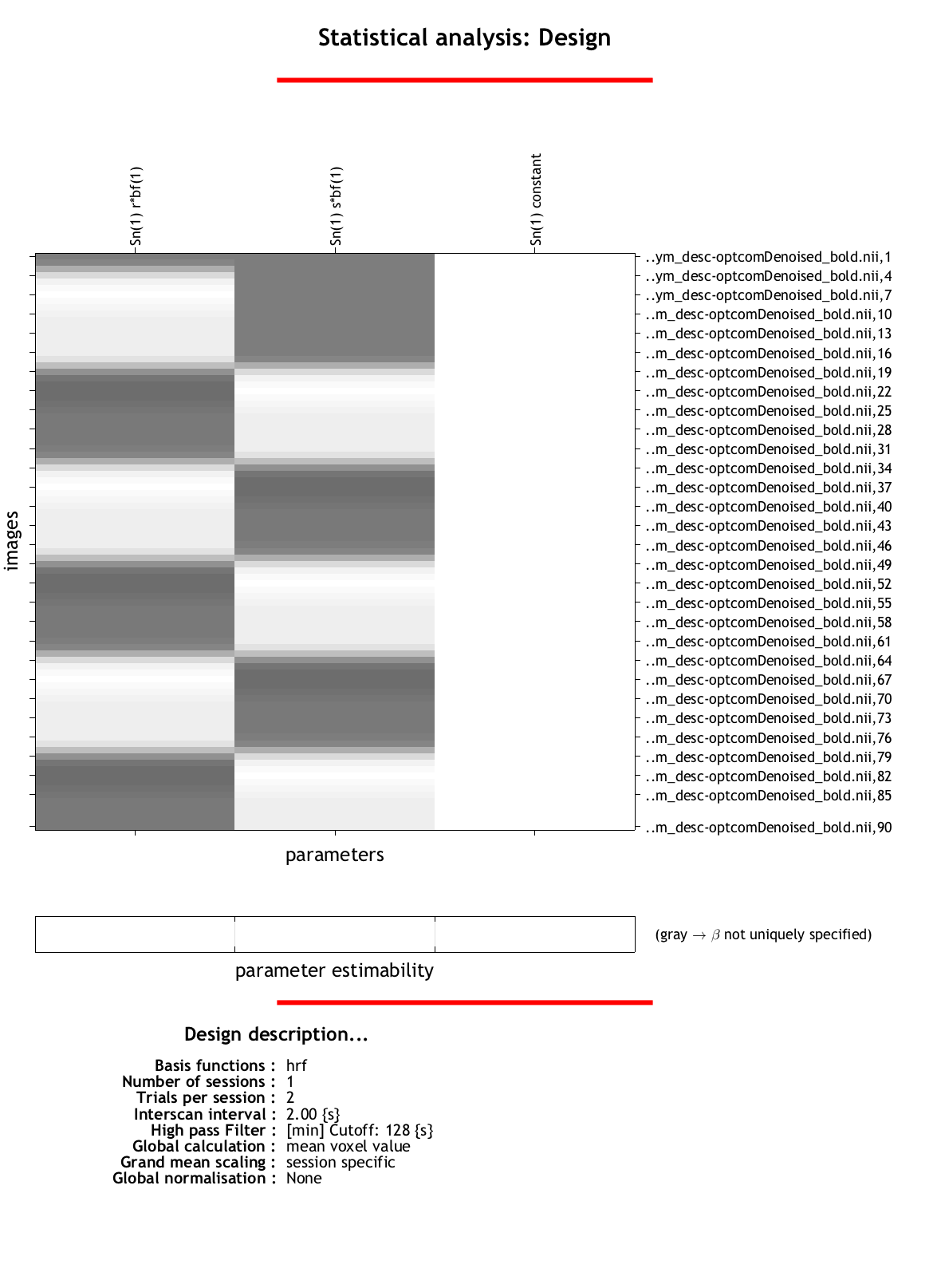
In addition, I think the task may not be optimally designed. Ideally you want to compare to some sort of baseline in the data - so you have task A, task B and then null periods of nothing at all. You also want the scanner to run longer after the last even so it can return to the non-HRF baseline - you can see that the 2nd column is white all the way to the end, as if the scanner stopped right at the end of the task - meaning the full HRF is chopped off.
All that said - you should still get an activation map, so something else is an issue.