Our team is performing QC on MRI data we have collected, and we came across some functional data that is a bit of an outlier. Across all three of our tasks, we are finding a large-intensity signal in the front of the field of view, outside the brain. The anatomical T1 seems perfectly normal, and we haven’t identified any consistent/major movement issues. I am wondering whether this is simply a strong aliasing artifact or whether it is something else too; we have only seen it on a few out of around 100 scans at the same center. We are on a Siemens scanner with a multiband acquisition.
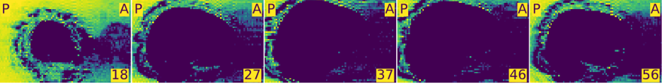
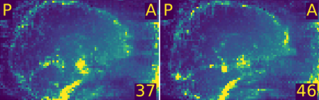
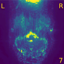
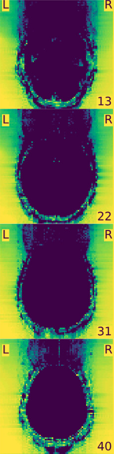
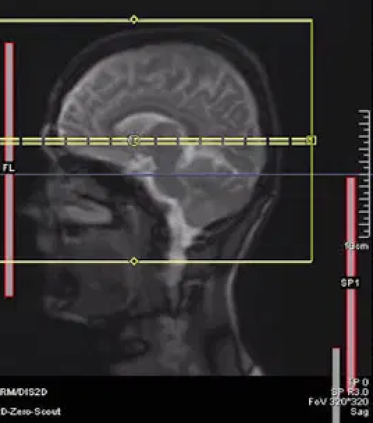
Below are some images that mriqc produces with heightened contrast to adequately show artifacts in the background of a scan.
Were the coil elements in the front of your receiving coil activated?
Was there some intensity bias correction done on these images before QC assessment?
What was the phase encoding direction? Was there some parallel acceleration (GRAPPA?).
I’m not sure about the coil elements, since this data was gathered some months ago and probably no one remembers (unless it is logged somewhere in the dicom header, which I doubt?).
This is from mriqc, so I don’t believe any intensity bias correction was done yet.
Phase encoding is A>P (json code j-); GRAPPA = yes, Acceleration Factor PE = 2
This is wrap around in the phase encoding direction. You can see this most clearly with the ghosting on sagittal slice 18. The easy fix would be to increase your field of view.
I was under the impression that for wraparound you have to see the back of the brain being cut off and that same cut-off bit would be present in front, but this is much more dramatic and the spot with the most intense signal is also more inferior than I would have expected. Maybe the wrapped-around bit also included damp hair or some sort of hairtie?
Since we won’t be re-collecting, I guess the field of view increase is moot.
@neurolabusc thank you! I checked in the json files and it looks like, unlike most of our participants who have the output you have written above, the few problem participants are missing the “HEA” part:
@jsein good catch. It looks like the anterior head coil elements (HEA) were not plugged in for all of these acquisitions. Siemens software will still acquire images without the anterior elements, but the image will be degraded. When you latch the anterior element of the 20-channel head-neck coil, or the plug for the 32/64 channel coils, make sure to check the monitor built into the MRI scanner to make sure it registers the anterior elements. You can also check this on the scanner console, by making sure that the desired coils are highlighted in red (in your case HEA and HEP):
Yes, it may be tricky to see on the magnitude images if the bias correction filter (“Prescan Normalise” or “Normalise” in Siemens ) is applied at the console. Indeed the signal in the front of the brain will be artificially enhanced to match the signal from the back of the brain which is close to the activated posterior coli elements. If you look carefully, you will see that the noise in the background in also increased in the frontal part of the image but you have to watch carefully to see it. In temporal standard deviation images as you show, it is much more visible.