Dear experts,
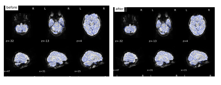
I ran fmriprep as part of the halfpipe pipeline, and I noticed that sometimes the before and after (moving) images of the sdc figure seem reversed. That is, in the after-image there is a gain of tissue in frontal and posterior areas etc. The AP and PA blibs seem fine. The input format is the same for everyone.
Do you have any idea what this means?
Thanks!
Best
Hans
Hi @hansvanderhorn,
Please post some images that shows how your images are before and after SDC is performed.
Best,
Jagan.
Hi Jimmy,
Thanks for your response. See below:
It seems like that before there is tissue gained relative to after. I checked all paths to the blibs, and they seem to be okay.
Best
Hans
Hi @hansvanderhorn ,
A few things come to mind.
- Check to ensure that the AP/PA EPIs you are using as “fmaps” do indeed have opposite PhaseEncodingDirections listed in their json files.
- Check that the PhaseEncodingDirection fields in the json of your functional BOLD and the corresponding directional EPI fmap are matching.
- You could try running the preprocessing again for this subject WITH and WITHOUT field map correction, and in both cases specify the T1w as one of the output spaces so that you can actually overlay the preprocessed bold output on to the anatomical and check how well they match. Ideally, the output with fieldmap correction should have a better functional-structural overlay. If you do try this, to save time please make sure to reuse the freesurfer output/directory it already generated for this subject.
What version of fMRIprep are you using? Based on discussions on here certain versions of fMRIprep didn’t do so well with certain SDC corrections.
Best,
Jagan.
Hi Jagan,
Thanks!
I checked the first two comments, and it all matches. De functional data has j-, the AP blib also j- and PA j.
I ran fmriprep within the halfpipe pipeline, but I’m not sure which version of fmriprep runs in it (I downloaded halfpipe recently).
Best
Hans
I found it, it’s version: 20.2.7 in halfpipe.
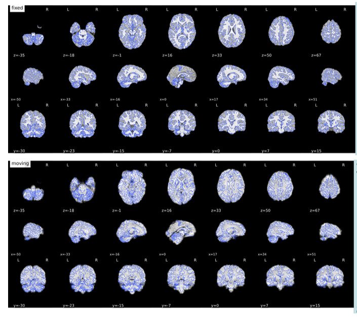
@hansvanderhorn, when you overlay the subjects functional (in T1w space) with the structural, does that look good?
Thanks for the suggestion, I’ll double check that first thing tomorrow. So far I think the alignment went well.
What I also noticed is that for some subjects the frontal and temporal ‘gain’ after correction is less, not very apparent, or sometimes it looks even like there is some tissue lost. However, what seems to be consistent for all subjects I checked so far, is that there is always some occipital and cerebellar/brain stem ‘reduction’. I also checked with some of my previously processed data where I used fmriprep’s fieldmap less correction, and I also noticed that consistent pattern, although the frontal areas always seem to gain tissue.
Long story short, it might be the case that this correction is going well after all (especially because all input seems to be right). I’m also curious of other peoples’ experiences.
They overlay looks fine in my opinion:
I guess the sdc did not go wrong after all. I was under the impression that sdc always results in a ‘gain’ of tissue (frontotemporal), but I guess that’s not the case.
Hi @hansvanderhorn ,
Glad to hear there are no issues.
Generally speaking, SDC correction should always improve the functional-structural alignment following coregistration. Since SDC’s goal is to correct spatially distorted/warped signal so that it can be “put” back to where it is supposed to be be, the best way to determine if it was successful is by comparing it to the structural (which in general hasminimal to no distortion).
Best,
Jagan.
Hi Jagan,
That makes sense. Thanks so much for all your help and explanations!
Best
Hans
1 Like