Hello all,
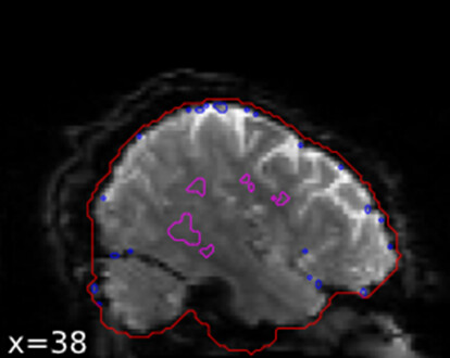
We are currently manually reviewing the derivatives output of fmriprep in a study of Alzheimer’s disease patients and healthy controls. I have noticed that here is a pattern in some of the sub-**_task-objectmemory_run-_desc-rois_bold.svg reports that I have seen thus far. The brain mask around the inferior portions of the brain seems to “droop” and includes some dark space around the MTL. I don’t know if this is maybe due to signal dropout in this region. Since our study is focusing on the MTL, we do not want to take any chances on data quality in this region.
Here is one example (new users are only allowed to embed 1 image at a time):
Any thoughts?
Thank you in advance!
– Ivan K
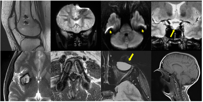
Seems like a clear case of a susceptibility artifact that occurs close to large air cavities. See this page for visual reference: Susceptibility artifact - Questions and Answers in MRI, see row 1 column 3 in the figure:
I guess the brain mask is derived from anatomical images which are less affected by the susceptibility artifact due to different image acquisition methods. One way of dealing with these artifacts is to perform a geometric distortion correction (e.g. FSL TOPUP: topup - FslWiki). I bet you will find similar distortions around the orbitofrontal cortex too.
Thank you for your response! I suspected susceptibility distortions as well. I will take a look through the fmriprep documentation and see which images the structural and functional masks are derived from and see if there is any additional correction that should be performed.
– Ivan
1 Like