We recently acquired functional scans for a project in which we’re interested in getting high resolution of brainstem and some subcortical structures.
We got a full, whole-brain anatomical image but for the functionals, we acquired only partial brain. We wanted to know whether fMRIprep works on this partial data, so I ran an initial test run on one of the functional datasets. It seemed to work fine (the anatomical is well aligned to the functional slab; compCor seems to have generated reasonably).
Is there something missing that might indicate fmriprep has not done a sufficient job? Is it able to run partial slabs? Any information on this would be immensely helpful! Thanks!
The short answer is yes, it seems that fmriprep is able to deal with partial brain slabs. Your example is a good example, we also got an example at our lab where the alignment between a partial FOV functional image (30 slices of 2mm along the Sylvian fissure at 3T) and a full FOV T1w image worked well.
If the alignment between your bold image and the T1w is good, I would say that you are good to go!
Additional question: Do you use SDC to correct your functional images?
There were some other discussions about this subject in those threads:
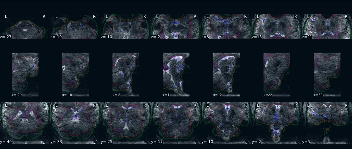
I have run into an issue with the partial brain functional mask. While the alignment of the functional and anatomical seems really good, the mask is coming out very misshapen:
I can’t tell if my AROMA-ICA components are all noise because the mask is unusable or if it’s because the protocol we are using isn’t feasible (we’re trying some experimental setups with high resolution).
Is there a workaround to this? Is there any software that can handle partial slabs for functional scans? Given the experimental nature of our protocol, we’re trying to figure out if we’re getting any good signal in the brainstem. Any advice or info would be incredibly helpful here! Thanks!
On this figure, I think that we looking at the masks for aCompCor, not AROMA-ICA. In red you have the brain mask which seems particularly off. In green is the edge mask which looks correct. In magenta you have the mask merging the WM+CSF compartment for anatomical CompCor ROI, excluding any GM partial volumes, which looks relatively ok (hard to see on the figure). In blue it is the temporal comCor ROI. The underlying image is indeed very noisy, it must be challenging to process it!
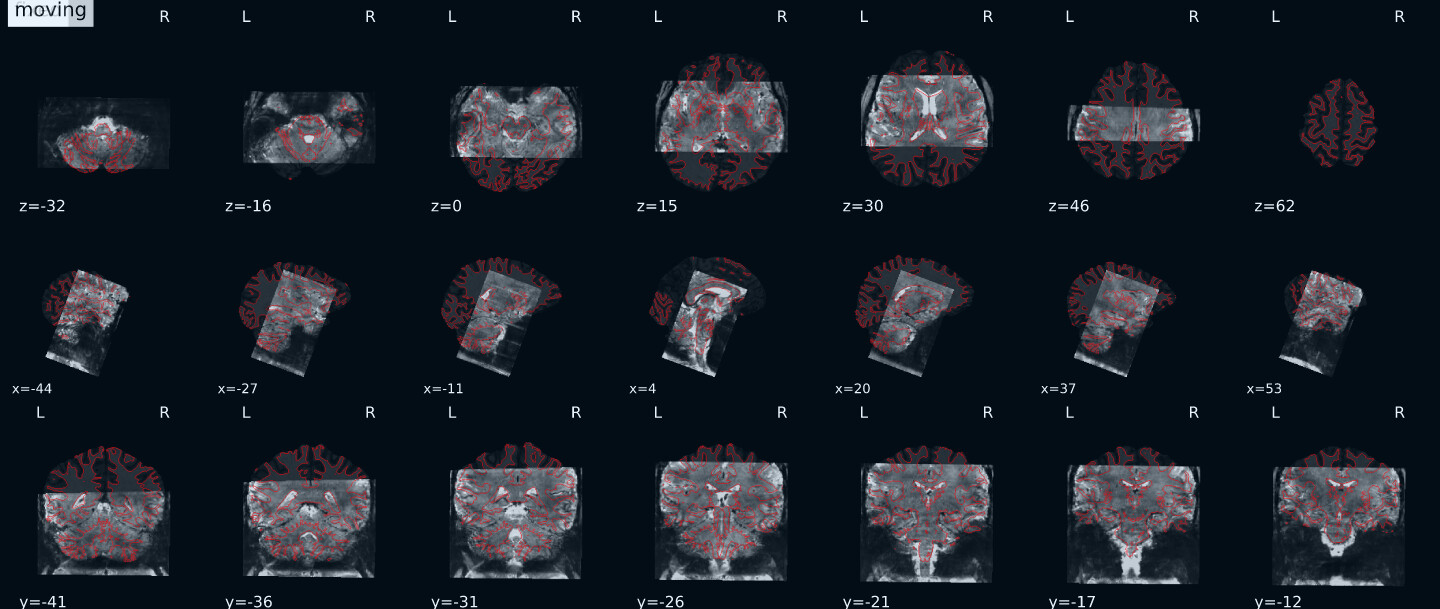
It could be interesting to see the image showing the anatomical and functional alignment to assess how well it went. Did fmriprep use any susceptibility distortion correction (SDC) in your case?
There’s been a problem with the distortion correction in fmriprep, though I believe this is due to some kind of naming issue or something trivial. I’m currently working on trying to get that sorted out so I can see if fmriprep, when using the field maps, can help with the noise. The thread for the fmap issue can be found here: Fmriprep TOPUP Issue Not Finding Files - #4 by Steven
Did you ever solve this issue? I am running fMRIPrep on slab data and I am encountering the same phenomenon that the brain masks for the functional data are terrible.