I have just run a subject through the default CPAC pipeline and got the following two quality images.
Does this look like a segmentation issue to you all? Or is this a typical output?
I have just run a subject through the default CPAC pipeline and got the following two quality images.
Does this look like a segmentation issue to you all? Or is this a typical output?
Hi @asd_tms, Thank you for your query.
Could you please answer following questions for me so that I can help you better?
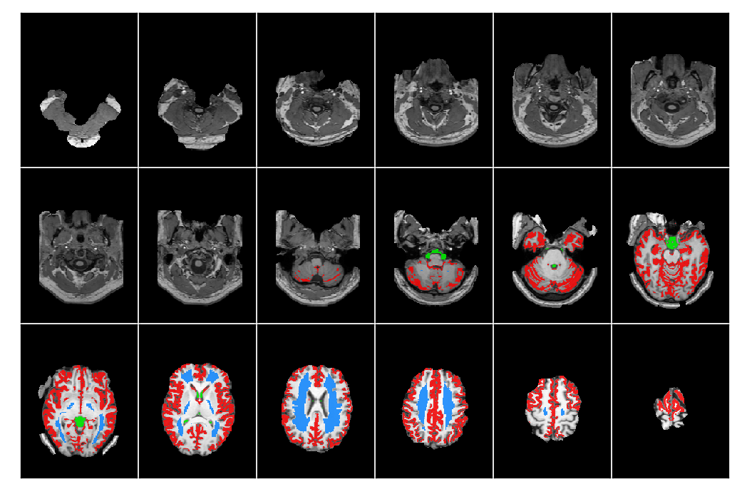
Hi:
Here are the outputs in the output directory, from my anat folder:
sub-BNI_ses-1_desc-brain_mask.nii.gzsub-BNI_ses-1_desc-head_T1w.nii.gz
sub-BNI_ses-1_desc-preproc_T1w.nii.gz
sub-BNI_ses-1_from-MNI152NLin6ASym_to-T1w_mode-image_desc-linear_xfm.nii.gz
sub-BNI_ses-1_from-MNI152NLin6ASym_to-T1w_mode-image_desc-nonlinear_xfm.nii.gz
sub-BNI_ses-1_from-MNI152NLin6ASym_to-T1w_mode-image_xfm.nii.gz
sub-BNI_ses-1_from-MNI152NLin6Sym_to-T1w_mode-image_desc-linear_xfm.nii.gz
sub-BNI_ses-1_from-MNI152NLin6Sym_to-T1w_mode-image_desc-nonlinear_xfm.nii.gz
sub-BNI_ses-1_from-MNI152NLin6Sym_to-T1w_mode-image_xfm.nii.gz
sub-BNI_ses-1_from-T1w_to-MNI152NLin6ASym_mode-image_desc-linear_xfm.nii.gz
sub-BNI_ses-1_from-T1w_to-MNI152NLin6ASym_mode-image_desc-nonlinear_xfm.nii.gz
sub-BNI_ses-1_from-T1w_to-MNI152NLin6ASym_mode-image_xfm.nii.gz
sub-BNI_ses-1_from-T1w_to-MNI152NLin6Sym_mode-image_desc-linear_xfm.nii.gz
sub-BNI_ses-1_from-T1w_to-MNI152NLin6Sym_mode-image_desc-nonlinear_xfm.nii.gz
sub-BNI_ses-1_from-T1w_to-MNI152NLin6Sym_mode-image_xfm.nii.gz
sub-BNI_ses-1_label-CSF_desc-preproc_mask.nii.gz
sub-BNI_ses-1_label-CSF_mask.nii.gz
sub-BNI_ses-1_label-CSF_probseg.nii.gz
sub-BNI_ses-1_label-GM_desc-preproc_mask.nii.gz
sub-BNI_ses-1_label-GM_mask.nii.gz
sub-BNI_ses-1_label-GM_probseg.nii.gz
sub-BNI_ses-1_label-WM_desc-preproc_mask.nii.gz
sub-BNI_ses-1_label-WM_mask.nii.gz
sub-BNI_ses-1_label-WM_probseg.nii.gz
sub-BNI_ses-1_space-MNI152NLin6ASym_desc-brain_mask.nii.gz
sub-BNI_ses-1_space-MNI152NLin6ASym_desc-head_T1w.nii.gz
sub-BNI_ses-1_space-MNI152NLin6ASym_desc-preproc_T1w.nii.gz
sub-BNI_ses-1_space-MNI152NLin6ASym_label-CSF_mask.nii.gz
sub-BNI_ses-1_space-MNI152NLin6ASym_label-GM_mask.nii.gz
sub-BNI_ses-1_space-MNI152NLin6ASym_label-WM_mask.nii.gz
Additionally, I get several .json files and the two images I included in the original post.
Thanks!
Thank you @asd_tms for your reply and further clarification.
Skull Stripping quality varies/depends on both tool and data so, I would recommend you to try few of the skullstripping options available, on a couple of subjects first, to compare the quality.
You can also fork these methods so that you get all outputs from different methods on a single run, which is helpful for comparison.
Would you mind sharing your pipeline.yml file also?
MANAGED BY INCF