Dear fMRIprep experts,
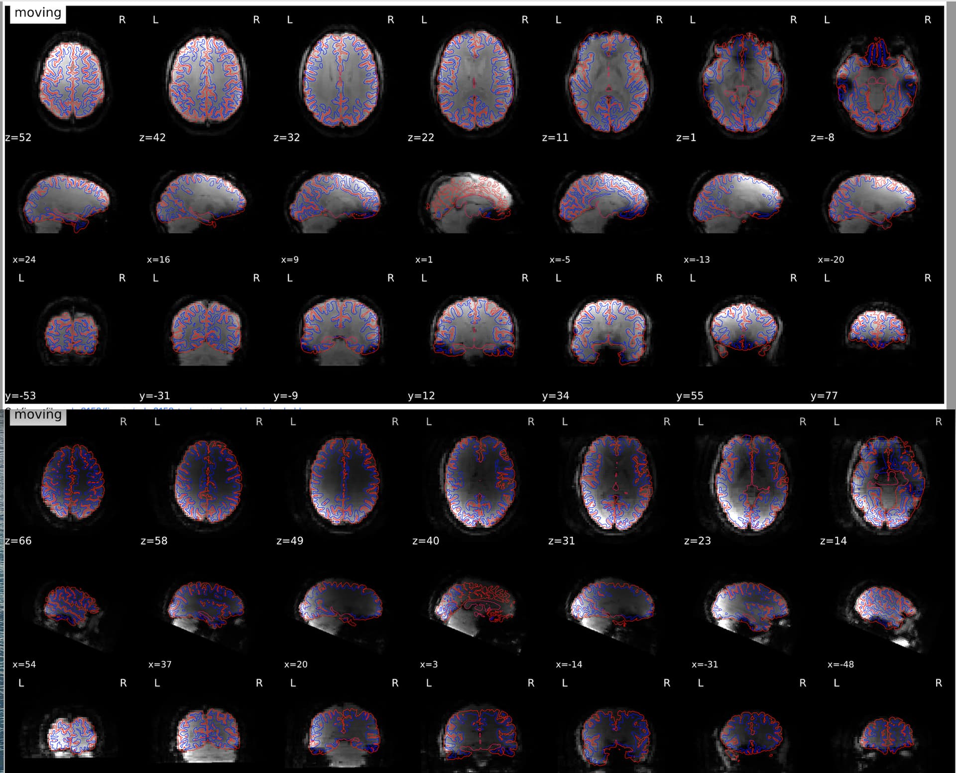
I am new to fMRIprep and noticed that dark images appeared under functional and anatomical alignment section for several participants despite normal raw scans (and normal maximum intensity values) based on visual inspection (a screenshot of two examples is provided below; lower one is more severe compared to the upper one). I was wondering what might be the potential causes of this and if it warrants exclusion of scans?
fmriprep version: 22.0.2 run on a HPC cluster
Command used: singularity run -B “{myproject}":"{myproject}” --cleanenv “{myproject}/fmriprep-22.0.2.simg" --skip-bids-validation "{myproject}/nina” “{myproject}/nina/nina_Processed" --mem_mb 30000 participant --participant-label "{SUBJECT}” --fs-license-file “${myproject}/license.txt” --output-spaces MNI152NLin6Asym:res-2 MNI152NLin2009cAsym:res-2 --use-aroma -w /tmp/
Type of SDC used: No SDC flag or fieldmap was used or included
Many thanks in advance for your assistance with this issue!