I have been looking into the difference between T1 and T1-weighted MRI, but I could not find any. Are there MRI images that show the differences between those two types of T1 MRI?
1 Like
Hello,
Those terms are synonymous.
Best,
Steven
2 Likes
Hi,
Out of curiosity: I thought that T1 can refer to quantitative T1 imaging, while T1w is explicitly non-quantitative (i.e. it reflects t1 contrast but the values cannot be taken as actual tissue T1). Am I wrong ?
Best,
Bertrand
Oh, that’s a good point, I suppose I didn’t consider qMRI. Usually I’ll see it written out as T1-relaxometry in that context. I guess it would have been better to ask first, @h_b, where have you seen T1 vs T1w come up in your readings?
I am dealing with two datasets; the first one is from this link and the second one is from this link. The second dataset description refers to the T1 as native. The second one (from their published paper) refers to it as T1W. When I compare two axial slices from two MRI images randomly selected from the two datasets, I see that the first dataset is more “clear” (i.e., shows more details/granular). The right and left figures belong to the first and second datasets, respectively. However, I am not sure if this is really because of the differences between native T1 and T1W images or if it is just different instruments (I remember reading that some of the MRI images in the second dataset were captured back in the 90s)
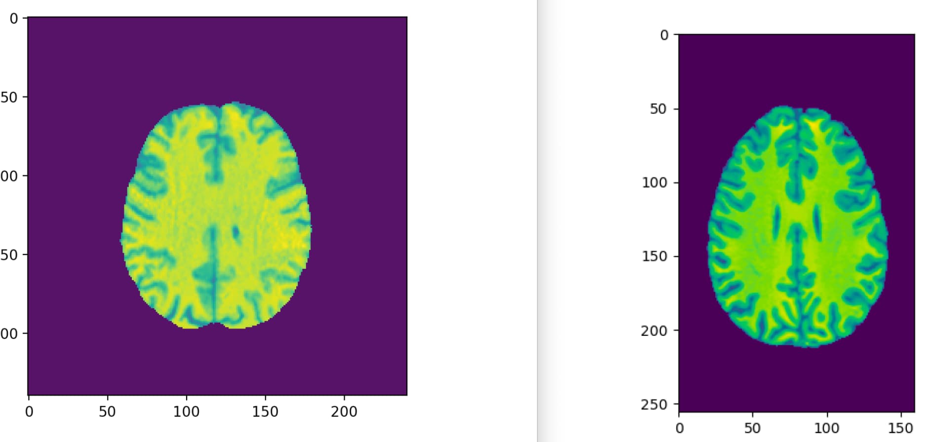
It looks like in the dataset associated with theUPenn link, they administer a Gadolinium contrast to enhance the T1 image (post-contrast, as opposed to native w/o contrast), but both are T1w images. At least that is my understanding.
1 Like
Their naming conventions call the Gadolinium contrast MRI images T1CE. What would be the reasons for such significant differences between the MRI images shown above?
You may want to look at the google slides from my lectures. T1 and T2 refer the two static contrasts that influence whether a tissue is dark or bright (there are also endogenous contrasts like diffusion or exogenous contrasts like a bolus of Gadolinium). A Steven noted, the terms T1, T1w and T1-weighted are all synonymous.
T1 effects dominate if both the echo time (TE) and repetition time (TR) are short, and T2 will dominate if both are long, while a long TR and short TE will balance these two effects (e.g. a proton density scan). Fat is bright in a T1 scan, Water is bright in a T2 scan, while a PD scan shows hydrogen concentration.
However, not all T1 scans look the same. One can choose an inversion pulse, use magnetization transfer pulses, enhance the tissue with an exogenous contrast (Gd), and other acquisition adjustments. One can also apply processing on the scanner or offline to adjust image inhomogeneity or smoothness.
When comparing different datasets, it is worth noting that different scanners have different capabilities and vendors have their own proprietary tricks that mean that data across studies can look different. Comparing data across field strengths (e.g. 1.5T vs 3.0T) is tricky as T1 (realignment) is proportionally slower with increasing field strength while T2 (dephasing) is faster.
3 Likes