Hi all,
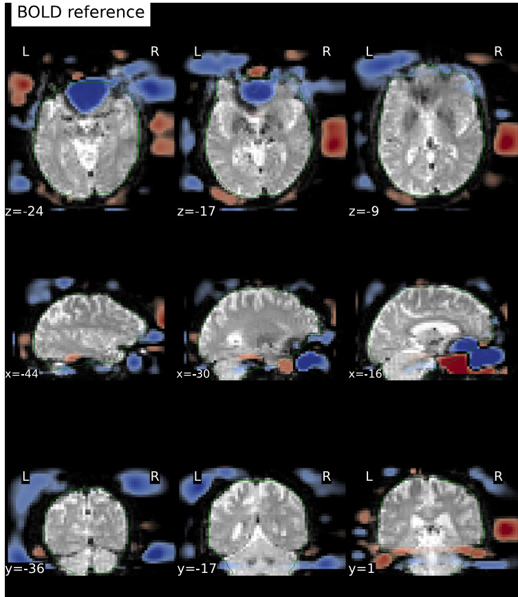
I’ve run fMRIprep 24.1.0 on ~ 150 datasets (PE-dir: func/RL_bold;fmap/LR_epi), and I noticed that for one of the subjects, the EPI and the reference fieldmap are not aligned well with each other.

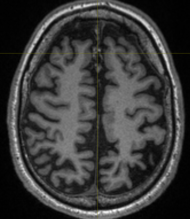
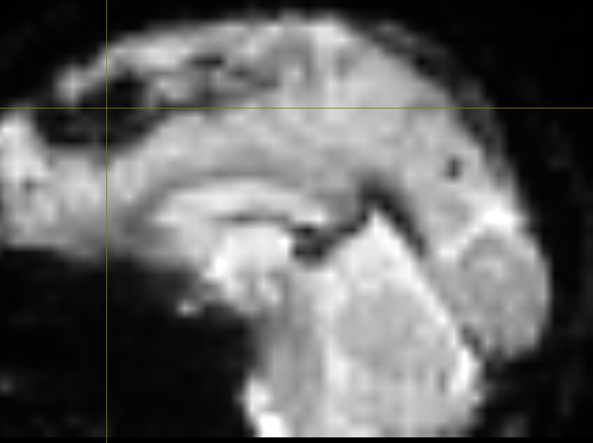
Upon further inspection, I noticed a midline “something” in the anatomical scan (below, anomaly marked by crosshair) extending several slices:
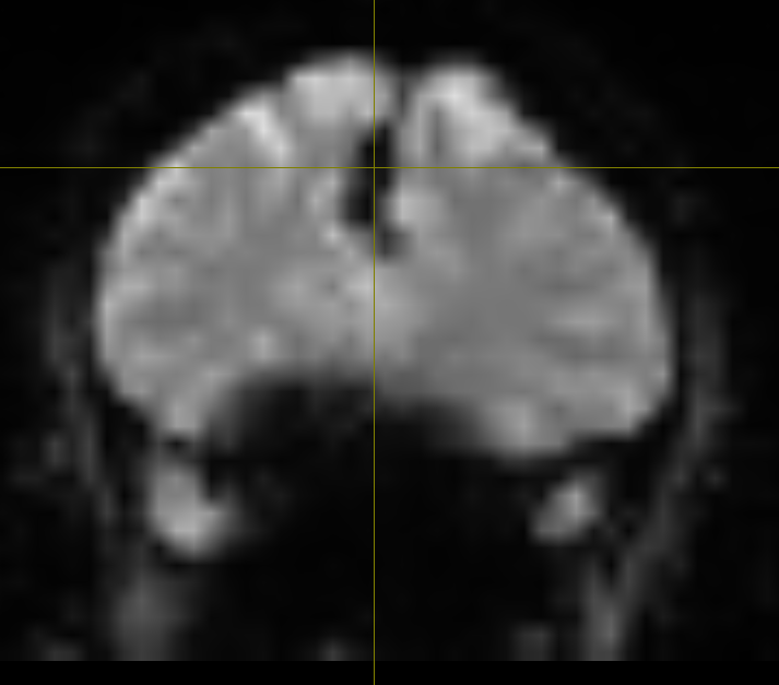
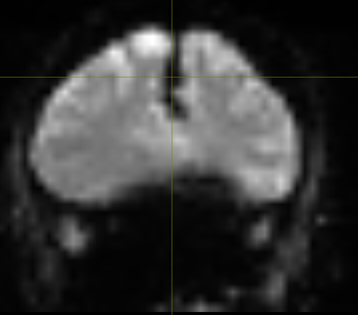
Which appears as a signal-void region, affecting LR_epi (one top) and RL_bold (two bottom) differently:
I would like to ask:
- By any chance, do you know what that is?
- What should I do to address this issue?
- Can it be caused because of that anomaly?
Looking forward to your comments!
Best,
Amir