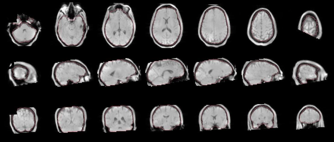
So I’m running fmriprep on some data from a pilot participant and, like a good researcher, I inspect the QA reports.
I notice that the skullstripping looks aggressive in the upper-right-most corner of the attached image, and suspect that there were some FOV problems during acquisition. However, when I load the raw field map (and bold!) data, there’s actually ample data in the z=70 slice and a couple of slices above that. Furthermore, the mask files that are output in the derivatives directory are more robust than the one shown and don’t have the abrupt truncation of brain.
So now I’m mostly just confused about whether this is a bug in the reports, a skull-stripping bug, or a bug in my own comprehension of things.
That is indeed weird. Have you looked at the preprocessed data for this? I suspect this is a report generation problem because if it were masking, the red contour would show the “bite” into the image.
The preprocessed data (in MNI space) is not truncated, so I agree that it’s likely a report generation issue. Is there any data people would be interested in seeing to help reproduce the bug?
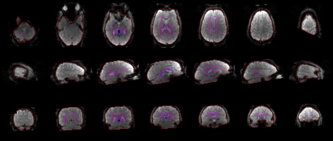
It’s actually even more replicable than that. I’ve run this subject twice – before and after adding the “IntendedFor” field in the field maps .json file, so one run had field map processing in fmriprep and the other run did not.
Attached is the image from the field map run showing the mask from the magnitude image as well as the image from the run without field maps showing the brain mask in EPI space.
(Also somewhat interesting – in the attached jpgs, you can see a black square in the lower left image of the two montages. This black square does not appear on the .svg version of the files. This could be a complete red herring from the conversion process, but also seems like it’s in kind of the right spot…)