Hello all,
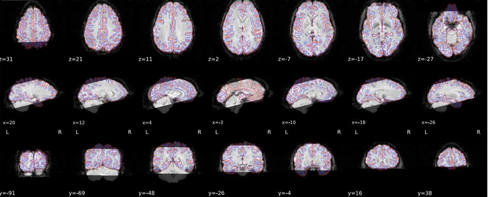
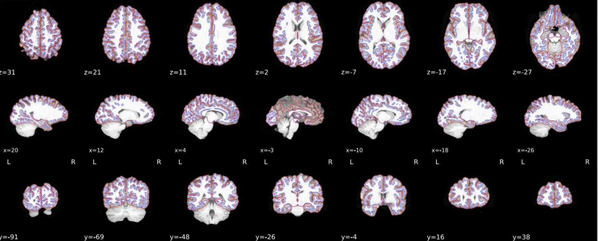
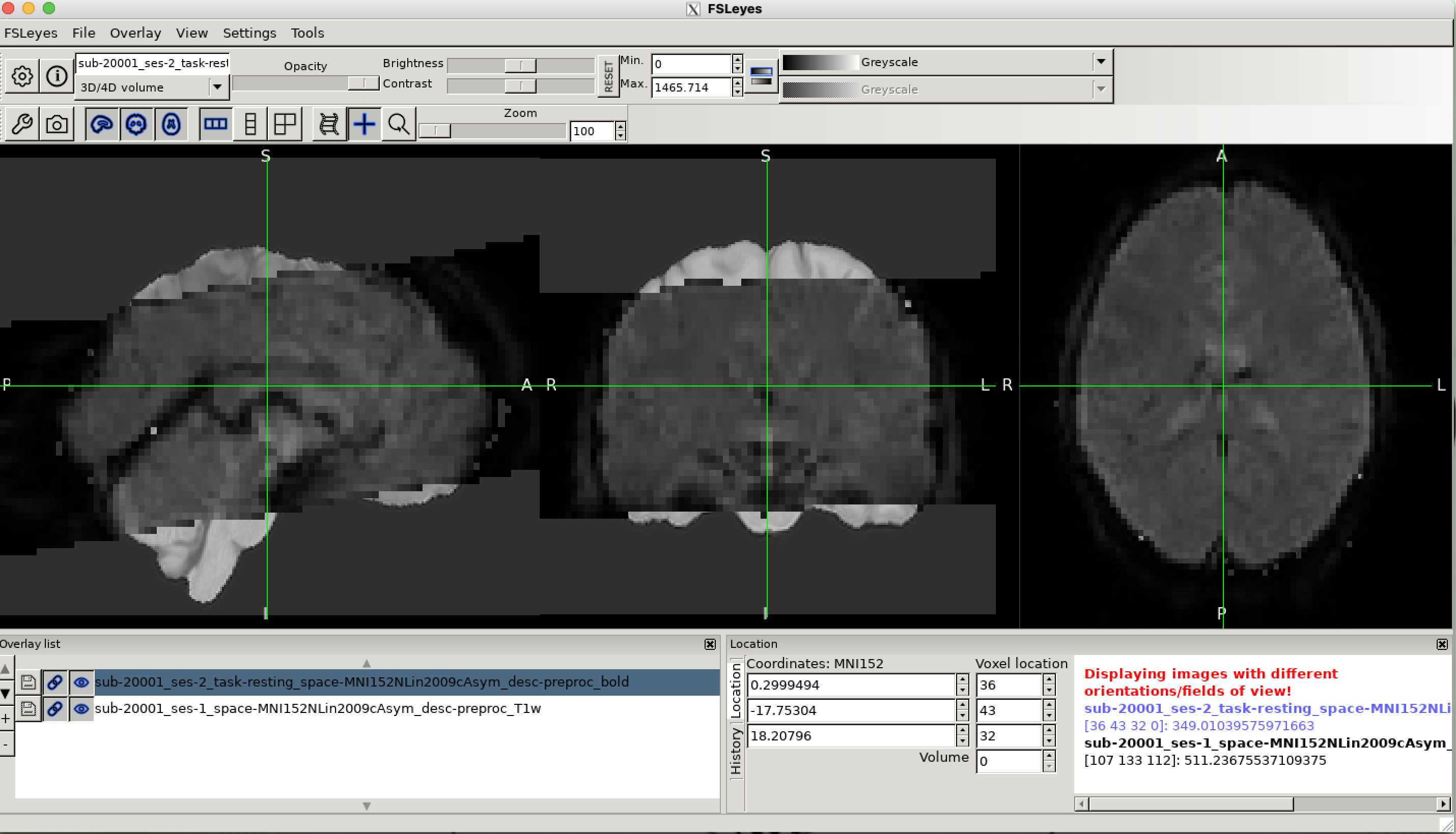
Grace again with another question. I successfully ran fMRIPrep on a couple of subjects. I was looking though the html outputs and they looked OK. The BOLD scans have a somewhat limited field of view (top cut off). However, when I look at the T1 and BOLD files in fsleyes I get the warning that the images are not in the same space.
I agree with you, it looks like the preprocessing went well, the functional image and the anatomical image look well aligned with each other.
You do not have to worry about the FSLeyes warning: indeed you are looking at two images that are in the same physical space, but their FOV and resolution are drastically different: one is your T1w image projected in the MNI space with a full FOV at the native resolution of your T1w acquisition while the other is your functional image with a partial FOV, also projected into the MNI space with the resolution of your functional acquisition.
Being in the same physical space means that the two brain images overlaps each other (if the preprocessing stages went well which is the case here hopefully) but in your case, the two images grids are different due to the differences in resolution and FOV of your two images displayed in FSLeyes.
To check that, you can click on the “i” on the top left of FSLeyes and look at the header information for each image and spot the differences in those informations.
If you wanted to have the same grid for both images (and thus not see the warning in FSLeyes), you could do “Tools > Resample image” in FSLeyes and resample ( = re-grid) your functional image in MNI space to your T1w image in the MNI space. But I would advise against that because that would make your functional image much bigger and for no use really, except for general comprehension.