Hi,
We are having some issues with the output of our ASLPrep. The software appears to be processing the scans but for some reason it’s outputting these huge holes in a subsection of scans (see below for a detailed explanation of the problem). I can’t seem to figure out why this is happening and some clues or if someone has had this issue before would be greatly appreciated. Full details below…
Data formatted according to a validatable standard? Please provide the output of the validator:
Passes BIDS validator in ASLPrep
Relevant log outputs (up to 20 lines):
ASLPrep finished successfully, no error reported.
Screenshots / relevant information:
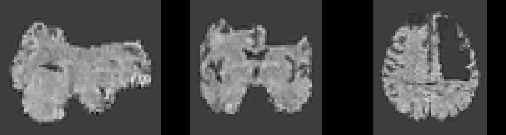
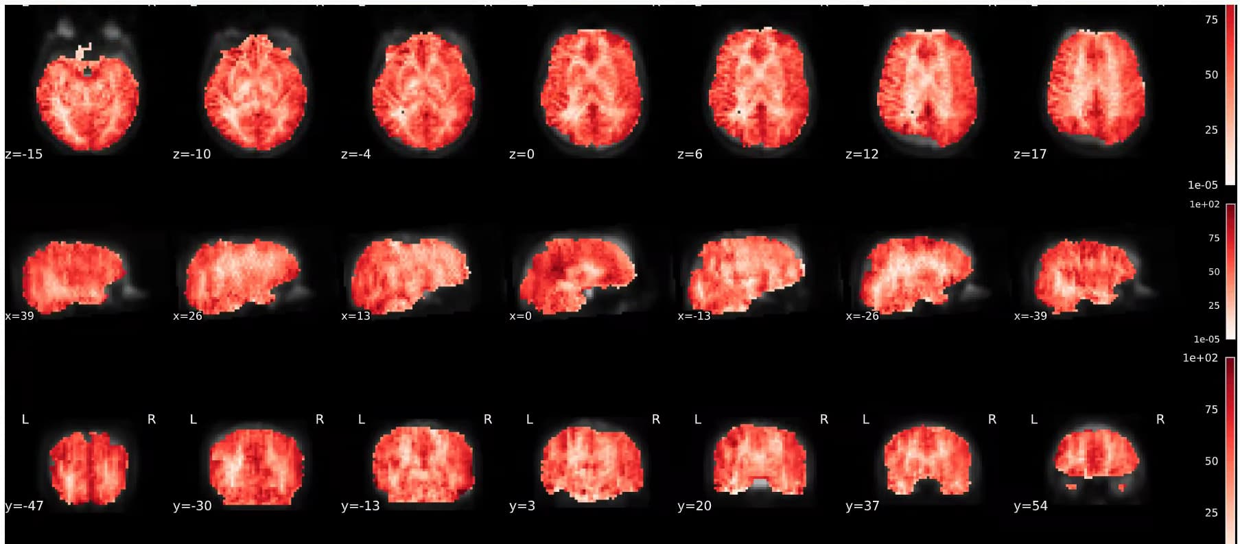
For some reason, they are cutting out random portions of our brains. Here are some examples of what is happening. This isn’t consistent across participants, but just appears to be happened on a subsection of participants.
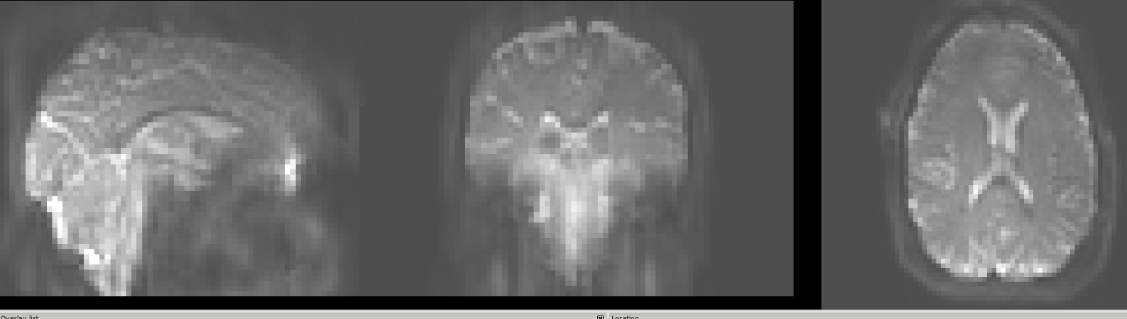
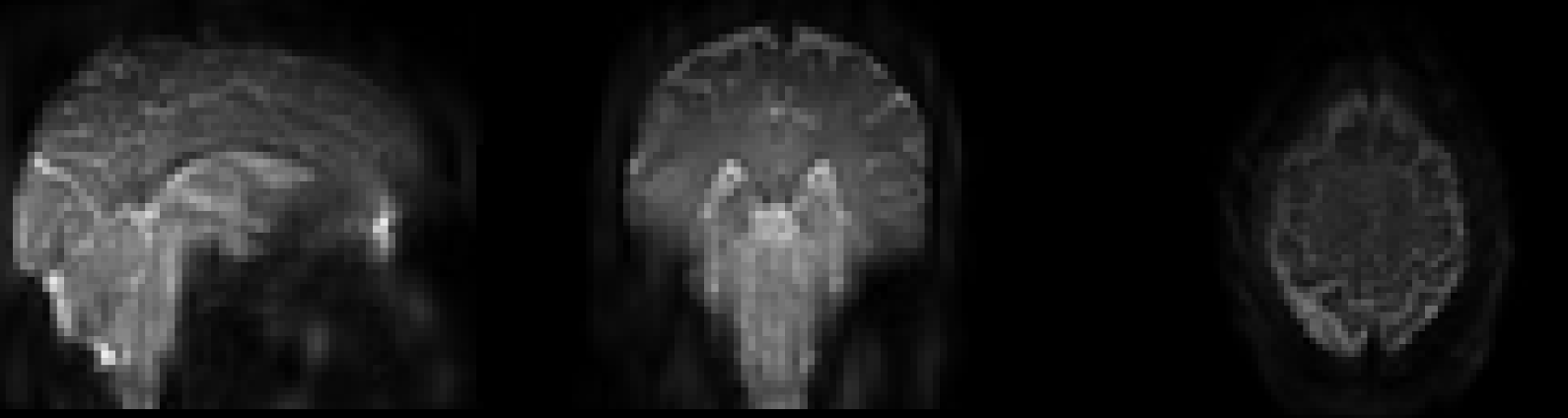
The alignment between the M0 and ASL perfusion before processing seems reasonable and there doesn’t appear to be any holes here? Here is the original perfusion image…
In the future, please use the Software Support post category and template, which prompts you for important information we can use to help debug your issue. You can see I edited your post for you this time. You can edit your post to add in the missing information.
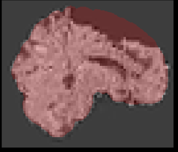
Beyond that information, I would also ask if rerunning on a fresh working directory helps. But it also seems like there is some pretty noticeable signal dropout in the top of the brain potentially leading to it being masked out.
Do passing subjects have the same kind of dropout?
Thanks, I edited as requested as much as possible. I also noted that I reran this in a fresh directory and still had this output problem. There does seem to be some minor signal drop out but this is pretty consistent across my ASL images and most of these don’t have this huge hole. My T1 mask does indicate that this portion of the brain should be included in the final perfusion output - so just wondering how I can go about fixing this? If it’s possible?
Hi @AustinBipolar. Any chance you could share the data from one of these problematic runs with me? I can try reproducing locally and see what step might be creating these holes.
Hi,
Sure what’s the easiest way to send. I’d like it only to be available to yourself. I’m not fantastic with how neurostars works. From what I could tell, the issue started AFTER MNI conversion step. The initial T1 looks bad but I substituted in a better one from another session and it didn’t seem to resolve.
Hi,
I’m facing the same issue — with multi-delay pCASL data, both the standard model and BASIL produce CBF maps with “holes” as if the ASL mask is too stringent. I’m using: