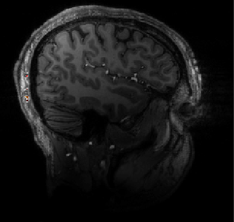
I’m working on fmri data preprocessing, and I’m facing this issue with T1w anatomicals having an intensity drift. As can be seen on the attached image, inferior slices have lower intensities and this poses issues in the segmentation step because tissue won’t be recognized correctly at the bottom brain slices. I was wondering if someone could recommend a method/software that can help me homogenize the intensity in my T1w anatomical images.
Another option would be to use AFNI’s 3dUnifize to stanardize+homogenize the brightness across the volume: https://afni.nimh.nih.gov/pub/dist/doc/htmldoc/programs/3dUnifize_sphx.html#ahelp-3dunifize
The program outputs an anatomical volume with WM scaled to an approx. uniform scaled value of 1000, and you can provide an option to try to scale the GM to an approximate uniform brightness, too. For example:
Thank you all for your helpful advice. I ended up using SPM’s segmentation workflow which did a relatively good job on my 7T anatomicals. I’ll keep your recommendations in mind for the future.