Hi all,
starting from the CT perfusion DICOM files I obtained, using 3DSlicer, the nifti penumbra VOI.
Now I want to check which anatomical areas are affected based on the AAL atlas (possibly using MRIcron, but other software are fine). I am stuck in the coregistration phase; I have already tried SPM without satisfactory results. How can I adapt a VOI extracted from a CT to an atlas?
Thanks for the advice, I am new to neuroimaging analysis, so any suggestions are welcome.
See the post here for how to do this in AFNI. Apply the computed transformation to move the region with 3dAllineate -1Dmatrix_apply. For ROIs, also use the “-final NN” option for nearest neighbor interpolation.
The Clinical Toolbox for SPM was designed for this purpose. It includes a tutorial with sample images to demonstrate how to use it. It is described here.
Hi everyone, and thanks for your advice.
I’m sorry for the confusion — in my previous post, I didn’t clearly explain the problem. I’m familiar with registering CT scans to standard spaces, usually after skull stripping (using 3D Slicer) and then processing with SPM or the integrated tool in DSI Studio.
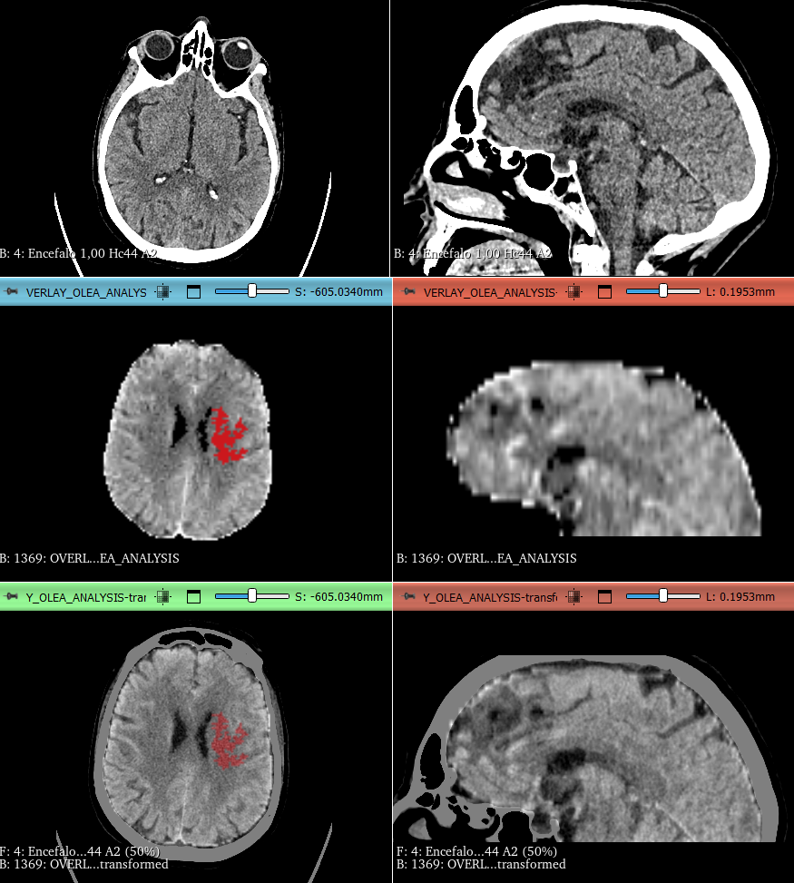
The issue is this: I have native brain scans and corresponding perfusion maps. The maps are generated from perfusion “raw” DICOM using Canon Olea software (I can also use Siemens Syngo.via, with similar results), but the output include only 31 slices and don’t fully cover the entire brain volume (see fig below).
While I can segment the maps in 3D Slicer and save them as a VOI volume, I’m unable to apply the same registration process to them as I do for the full brain scan.
I did not try AFNI, if possible I’d like to use only Matlab SPM, 3DSlicer, MRIcron and DSI studio; but if with this tools is not possible to apply ROI/VOI registration from native to standard space I will try AFNI.
Sorry for the difficulty but I repeat that I’m new to imaging analysis, I’m actually a medical resident.