I found it is very hard to find tools to remove skulls from CT images. All algorithms are made for MRI images. Tools are such as BET, BET2, 3DSkullstriper, ROBEX do not work with CT. Why are big different between both types of medical imaging. Also are there any CT skull removing available?
1 Like
I think there are numerous reasons why there are more tools for brain extraction of MRI versus CT.
- CT exposes participants to ionizing radiation which makes it less desirable for research in healthy adults. Most neuroimaging tool groups focus on healthy individuals. Even for research into disorders like autism, it is easier to get IRB for MRI versus CT.
- Clinical CT scans are often tuned for clinical questions, and are not ideal for brain extraction. For example, it is common to have numerous CT slices near the brain stem (where small injuries are life threatening) and very few slices for the cortex. These thick slices near the cortex do not yield good brain extraction. In contrast, slice thickness influences MRI SNR, so it is typical to have equidistant thin slices in MRI. This distinction is changing with modern CT systems, but it is still a factor.
- (Hard) bone has very little signal on most MRI modalities (it has less hydrogen than other tissues). Therefore, it does not tend to introduce partial volume effects. In contrast, for CT bone is the dominant contrast: bone has a Hounsfield of ~1000, whereas the whole spectrum of CSF, gray matter and white matter ranges from ~0…40 (though note Gd injections can influence blood). This huge contrast difference means tends to mean that CT brain extraction yields an unsightly ring around the brain.
This is unfortunate, as CT is the work horse of the clinic - fast, relatively inexpensive, few contraindications, etc. Good tools in this domain can have huge clinical impact.
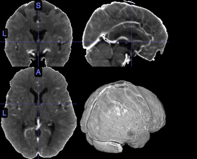
However, since CT intensity is calibrated, brain extraction is pretty easy. Below is a simple example
- I used a high resolution isotropic CT scan.
- I normalized the CT scan using the Clinical Toolbox for SPM. I suspect you could get a good result with FSL’s FLIRT using a good cost function.
- I thresholded the image so voxels darker than zero and brighter than 200 were set to zero. I used nii_thresh for this, but FSLmaths would work.
- To remove partial volume effects at the scalp-air boundary I ran FSL’s BET with ‘-f 0.1’
2 Likes