Hi community,
I am writing to share a significant update regarding the “Spontaneous Neuroelectrical Cascade (SNC)” hypothesis I previously discussed on this forum.
1. The Missing Link: The Mechanical Trigger
In my previous threads, I proposed a large-scale bioelectrical phenomenon but lacked a defined initiation mechanism. I have now successfully identified and documented the physical trigger: Voluntary Intracranial Pressure (ICP) Modulation.
- Previous Hypothesis (The “Event”):
2. New Evidence: The “Switch”
I have documented (via video and high-res photography) a voluntary maneuver—selective jugular venous occlusion without thoracic strain—that allows for the graded titration of ICP.
- Mechanism: This maneuver elevates ICP to a specific physiological threshold.
- Causality: Once this pressure threshold is reached, it acts as a mechanotransduction trigger, precipitating the SNC event described in the links above. The “Cascade” functions as a homeostatic release in response to the “Pressure.”
3. Physiological Markers & Question
During activation, I observe distinct markers: prominent Jugular Venous Distension (JVD) and immediate heart rate spikes/bounding pulses (confirming sympathetic engagement), which cease instantly upon release.
My Question for the Community:
To further bridge this gap between the Mechanical Input (ICP) and the Neural Output (SNC), what accessible sensors would you recommend for a home-lab setup?
- Would a standard SpO2 waveform be sufficient to document the hemodynamic coupling?
- Are there specific EEG montages (OpenBCI) best suited for detecting such large-scale “discharge” events triggered by pressure?
I have organized the visual evidence on OSF (currently private for review). Any advice on instrumentation to validate this Pressure-to-Cascade link is appreciated.
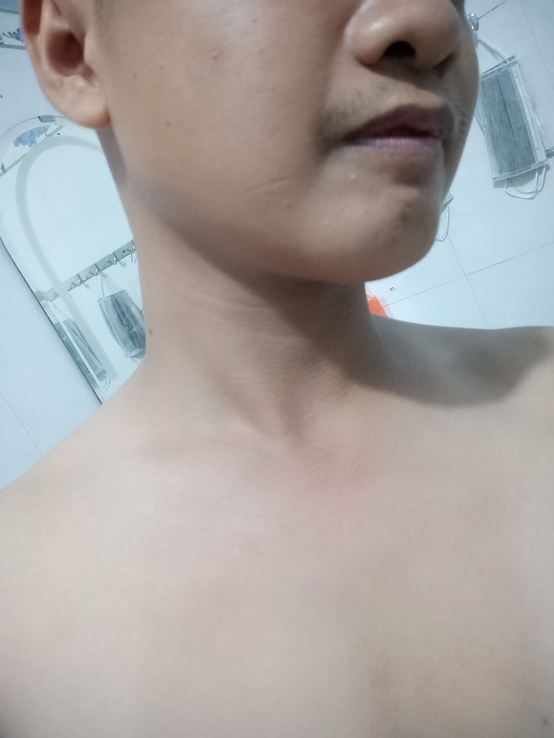
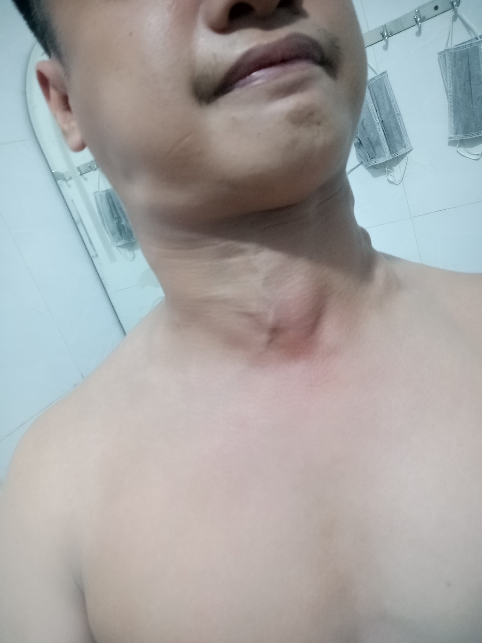
Figure 1. Visual Documentation of the Mechanical Trigger (Voluntary Venous Occlusion)
Image 1: * (Baseline State): The subject at rest. Note the normal anatomical profile of the cervical region with no visible venous distension. The sternocleidomastoid muscle and supraclavicular fossa are relaxed.
Image 2: * (Activated State): The subject performing the selective venous occlusion maneuver.
-
- Observation: There is a distinct and prominent Jugular Venous Distension (JVD) of the External Jugular Vein (arrow implied).
- Key Feature: Importantly, the pectoral region remains neutral (no chest heaving), indicating thoracic isolation. This distinguishes the maneuver from a standard Valsalva, confirming a localized blockade of venous outflow to elevate Intracranial Pressure (ICP).
Link to Video (DOI)
( ⚠️ STRICT MEDICAL DISCLAIMER & SAFETY WARNING
WARNING: DO NOT ATTEMPT TO REPLICATE.
The following post and associated datasets document a unique and highly specific physiological phenomenon for scientific observation and research purposes only. This is NOT a tutorial, a health recommendation, or a biohacking guide.
The maneuver described—involving the voluntary modulation of Intracranial Pressure (ICP) and selective Jugular Venous Occlusion—carries extreme physiological risks. Attempting to manipulate cerebral hemodynamics or occlude venous outflow without professional medical supervision can lead to:
- Acute Ischemic or Hemorrhagic Stroke: Due to sudden surges in intracranial pressure or disruption of cerebral blood flow.
- Permanent Neurological Damage: From localized hypoxia or mechanical strain on neural tissues.
- Retinal Damage or Vision Loss: Resulting from acute pressure spikes in the ocular system.
- Cardiac Arrest or Severe Arrhythmias: Triggered by intense autonomic reflex responses.
The subject in this documentation demonstrates a rare level of isolated motor control (thoracic relaxation during cervical occlusion) that may not be present in the general population. Any attempt to replicate these actions is done at your own extreme risk. The author and the platform disclaim any liability for injury or death resulting from the misuse of this information.
FOR SCIENTIFIC INQUIRY ONLY.)