Hi everyone,
I’ve run MRIQC via Docker (nipreps/mriqc:latest; v25.1.0.dev45+g699d1691) on a DWI dataset which has been acquired on a Siemens Prisma 3T and a Philips Achieva 3T scanner.
When inspecting the DWI-HTML-reports I noticed that the voxel-wise average maps show a reversed scale (Philips bright brain, dark background; Siemens: dark brain, bright background; see below), which was consistent across all shells and participants. I’ve also acquired resting-state fMRI scans. Here the scaling is not reversed in the voxel-wise average of the BOLD timeseries.
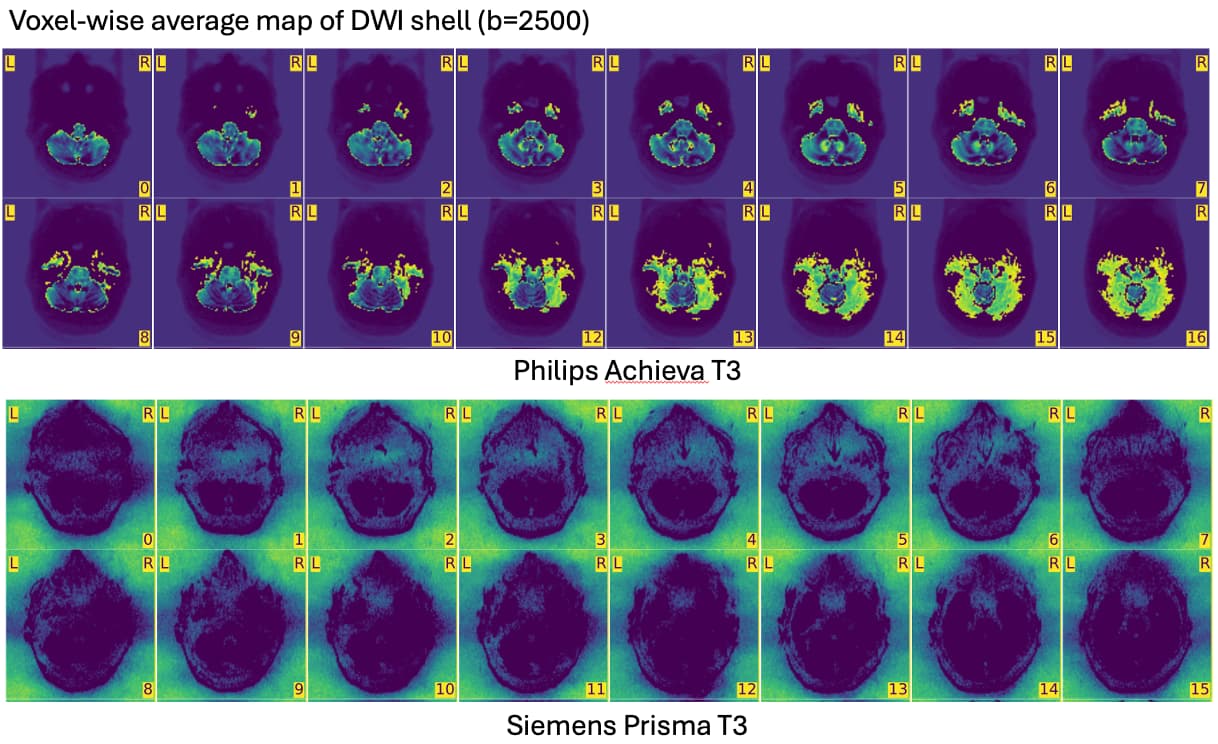
Consequently QC-metrics which are based on the background values (e.g. FBER) show a huge difference when comparing the two scanner sites. When checking the raw DICOM and NIfTI data, I noticed that in the Philips datasets, the signal intensity outside the skull was 0, whereas in the Siemens datasets, it was ~300 in the b=0 volumes.
I’m wondering if someone else has experienced this behaviour of MRIQC when using different scanners and how I should proceed the quality assessment of my dataset.
Thank you in advance!
Lionel