Dear All,
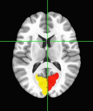
I am new to this field. Currently I am learning about parcellation of the brain using AFNI. I used “MNI152.NlinAsym09c.Brain.nii” provided by CarTool (1x1x1mm) as its underlay. For its overlay, I used two masks created separately using “3dcalc” (one for VisCent and onother for VisPeri) from “tpl-MNI152NLin2009cAsym_res-01_atlas-Schaefer2018_desc-1000Parcels17Networks_dseg.nii.gz” with the .tsv file provided by TemplateFlow. When I visualized the VisCent and VisPeri masks in AFNI, the pictures are seemingly swapped. That is, up to my current understanding, the VisCent-mask picture seem to show the peripheral visual (Lat Vis in Gordon et al 2017), and the VisPeri-mask picture does for central visual (Med VIs in Gordon et al 2017).
Do I understand the ROIs of the peripheral and central visual incorrectly?
Best,
Attha
P.S. I can upload only one picture. I choose the VisPeri-mask picture.
