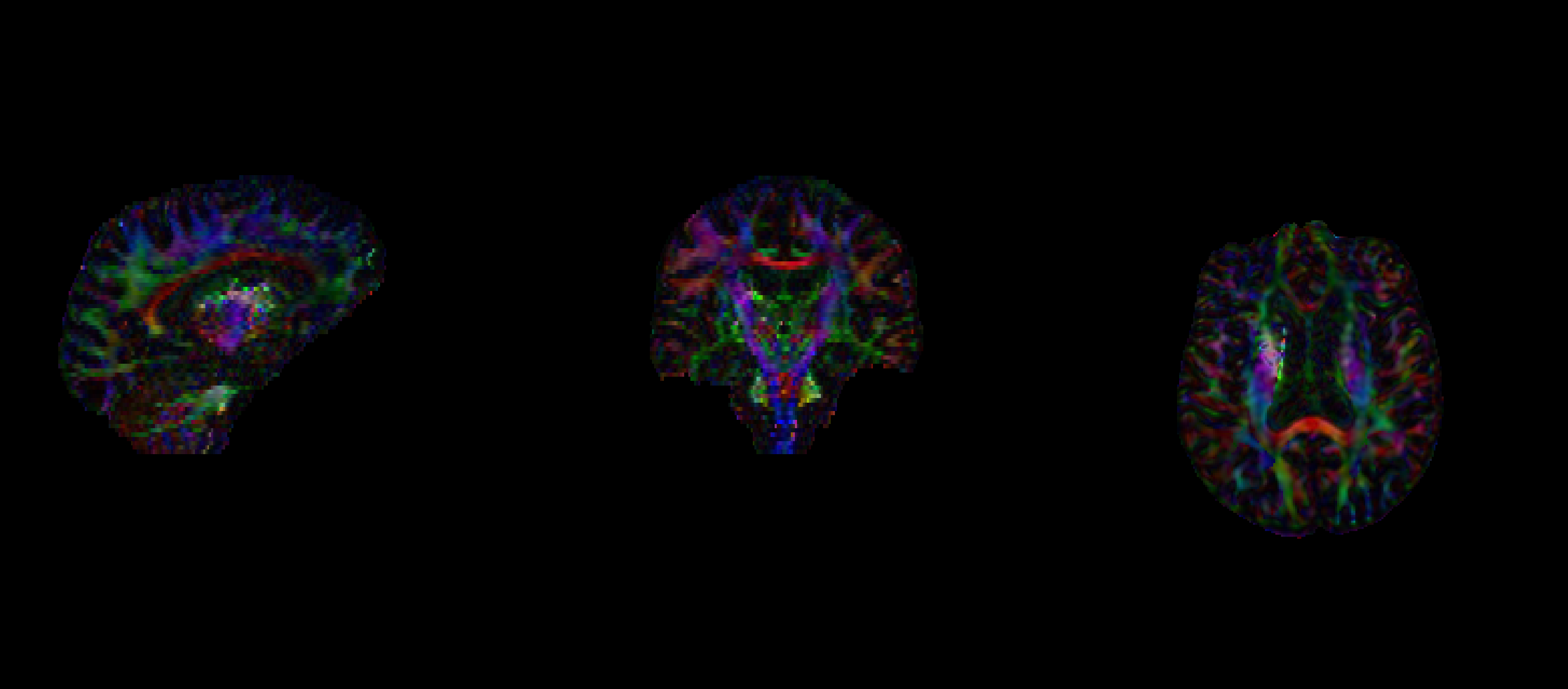
I’m relatively new to working with DWI/DTI images, and while reviewing my FA maps, I noticed what looks like an abnormality or possible artifact. I’ve attached an image below, there’s a bright spot that doesn’t look anatomical or expected.
I’m not sure if this could be due to preprocessing, acquisition artifacts, or something else. Has anyone seen something like this before? I’d really appreciate any advice or suggestions on what might be going on.
Hi @steven.jerjian
thanks so much for your reply.
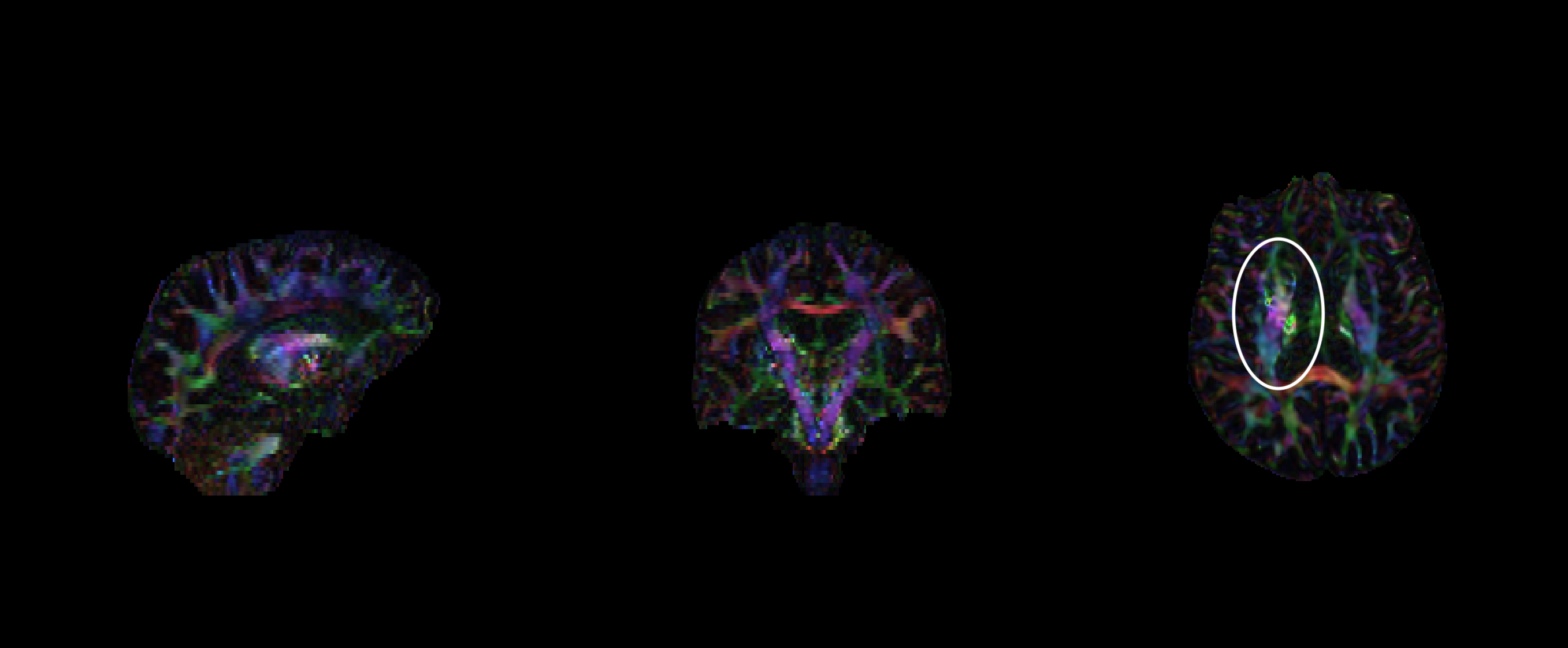
Actually, no, I mean the part I have identified in the attached images.
regarding my preprocessing, here are the steps:
1-denoising (dwidenoise)
2- unringing (mrdegibbs)
3- bias correction (dwibiascorrect)
4- dilated brain mask creation
5- susceptibility distortion correction (topup)
6- eddy current correction
7- bias correction (dwibiascorrect)
8- brain mask creation
9- Tensor fitting (dtifit)
Hi @RoSha, there may be something in your raw data, or the artifact may have been introduced by one of your steps. So rather than looking at the very final output of your pipeline, you should work your way through from start to finish, starting from the raw data, and checking the outputs of every step.
Hi @paulmccarthy .
thanks for the reply.
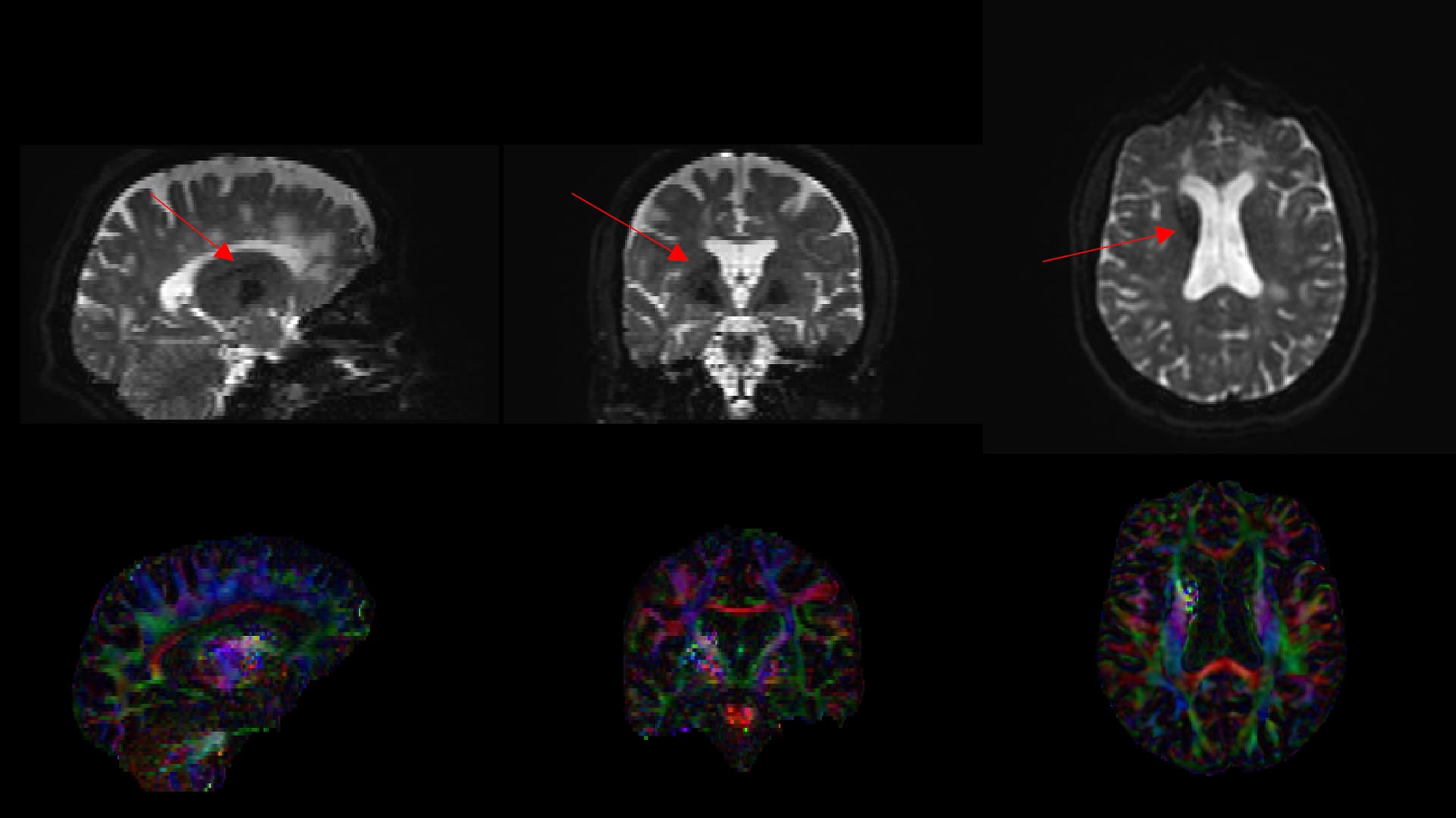
Actually what now I’m suspicious of is a black line that can be seen on the raw data (b0 image).
Do you think that could be the source of this artifact? if so, do you have any idea
what could be the cause of that black line, and how it actually can create such abnormality after fitting tensors?