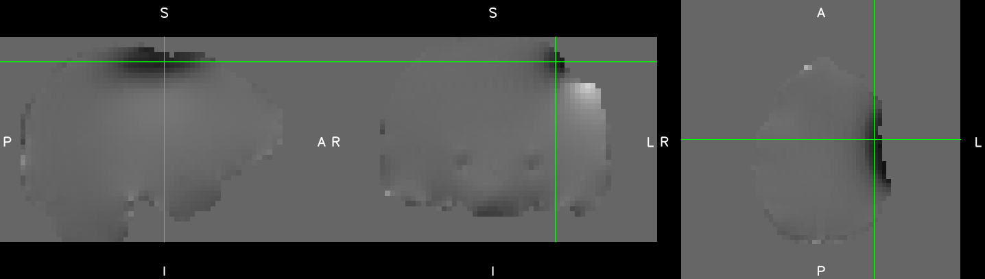
I have a question regarding susceptibility correction of BOLD EPI sequences with fMRIPrep and FUGUE (I acquired double-echo gradient echo images on a Siemens scanner) of patients that have deep brain stimulation electrodes implanted. My question is if FUGUE is a suitable algorithm for this, as the fieldmap is different from a “standard” one. Notably, there are some distortions due to the electrodes themselves, but also from coil wirings under skull near the motor cortex on one hemisphere, which cause a large area with signal loss but also with high values of signal distortion. I have attached a picture of an exemplary fieldmap.
I’ve read the original paper of fieldmapping by Jezzard and related work and to me there seems to be no mathematical reason why these fieldmaps should not work, or am I missing a crucial part here?
To me the big issue there would be the signal loss in those area due to the reduces T2* near the metal implant. The DeltaB0 field estimation in those area would be totally wrong as no reliable phase difference will be measured in those area with signal loss. If you may, you could share the magnitude and phase image of one gradient echo image that was used to generate this fieldmap.
In addition to the short T2*, there will obviously be strong geometric distortion on your BOLD image in this area near the metal implant. If possible, you should choose the phase encoding direction where there is more stretched area than compressed area, as it is easier for the algorithms to recover the signal from a voxel stretched over several voxels rather than recovering the signal from a voxel where the signal from several voxels has been piled.
There is also another option in FMRIPREP which is the “experimental” fieldmap-less approach, maybe you could try that as well. Basically I think this approach is doing a non-linear registration from the bold image to the T1w image. Maybe in you case it would work better than the fieldmap approach.
Your EPI images will be very affected by the presence of those implant, I am curious about the step you will use to mitigate the T2* and susceptibility effects due to the implant. My recommendation would be to go for short echo-time or multi-echo sequence, short echo spacing (through the use of high bandwidth and parallel acceleration). I didn’t look at the literature, perhaps also using non-EPI sequence for fMRI in that case would help?
Hi @jsein! Thanks for your quick and helpful reply!
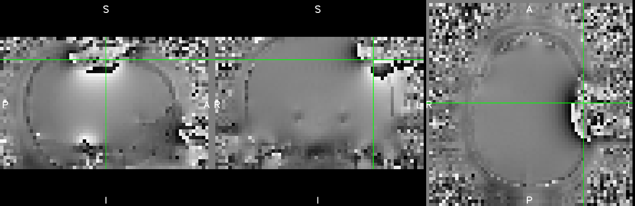
I attached a slice for the magnitude and phasedifference image. As you suggested, there is severe signal loss in the area where the wires are coiled beneath the skull and also around the electrode tips.
Thanks for the excellent I guess I would have to acquire separate EPIs with different phase encoding direction and compare them to judge, which areas get stretched and which compressed? Or is there another way how to detect this?
I’ve not thought about this but will try it out, thanks!
We are scanning at 1.5T with TE at 40ms currently. Do you think it makes sense to go shorter? Multi-echo is not an option I am afraid, as the stimulators are most likely not MR-conditional for these sequences (afaik nobody has done it so far and there is always a risk involved). Once I am at the scanner I would try different echo spacings with a higher bandwidth. We are aiming for TRs not longer than 2.7s, whole brain coverage with ~32 slices in a 3x3x3.3mm resolution.
What do you think about masking out the area with signal loss in the magnitude and phasediff images to prevent fMRIprep from correcting in that area? We would not use this area in the analyses anyway…
Yes masking the area of the fieldmap where the signal is lost and the phase difference is meaningless before unwarping seems like a good idea.
The same strategy hold for the non linear registration toward the anatomy, I saw paper doing so for the normalisation step for brain in the presence of brain lesions with ANTs software.(Analysis of automated methods for spatial normalization of lesioned brains - ScienceDirect)
But a good starting point would be to look at the epi images directly. I never saw one epi image in the presence of such strong susceptibility material, i wonder if there would be usable at all?
You are right there are safety issue as well to take into account, regarding heating as well as induced currents in the electrode. You must be careful then when thinking about increasing the bandwidth. You could look at different phase encoding direction and also different slice orientation (axial, coronal sagittal) to see where the artefact are less problematic…
Challenging experiment!