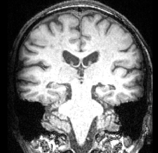
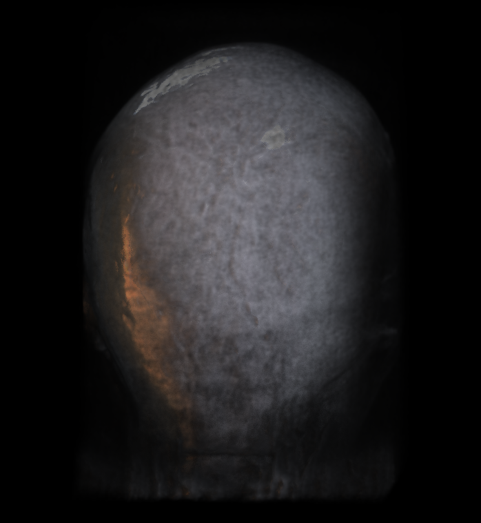
I came across one T1 image, in which the left side had apparent lower intensity than the right side of the image (the scalp is almost invisible at the left side), as shown in the figure below. I wonder what’s the potential cause of this kind of signal dropout. I think it is a hardware problem. Furthermore, this kind of data could still be used for valid analysis? I tried FreeSurfer and FreeSurfer faild to find the pial surface in these affected areas. Thanks for any help!
This data was from a legacy dataset, so that details about the acquisition may be not available (I mean I could not ask for help from the colleague who scanned the data). As far as I know, this T1 image was scanned using a 3-T Siemens scanner.
Many thanks! Another thing I am concerned with is that this kind of data is still usable? It seems that although the scalp is affected, the nearby gray matter regions seem less affected. If your guess is right, the gray matter signal is affected as well? If these regions were also affected, other parts of brain could be analysed? for instance, white matter and subcortical regions?
Yes, at first sight the brain image seems usable after correction of the intensity bias. It looks like you still have image contrast between WM and GM in the area of the dropout and the pial limit seems visible as well.
Do you have any suggestions about how to deal with the intensity bias? I tried FreeSurfer (v6.0) and based on the surface construction result, the pial surface is not extending far enough into the real boundary between gray matter and CSF. However, this kind of failure was also observed in the seemingly unaffected hemishphere (the other side of the image). So maybe this kind of failure is not related to the signal bias in my image.
I tried FreeSurfer 7.4, which used ANTs for bias correction denoising. Unfortunately, the result seems unchanged. So maybe more methods should be tried.
you can tweak the bias correction software to get a better intensity bias correction. Sometimes these software require a brain mask, you may have to optimize the brain mask creation to be sure to include in the brain mask the area of the brain with low intensity.