We would love some input on a couple of problems that came up during QC of our diffusion data, preprocessed in QSIPrep. All images here are taken from dmriprepviewer. I posted this in 5 parts, as I was only able to attach 1 image per post.
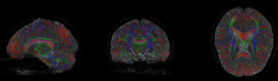
Part 1: We are noticing this red haze appears in many of our images, some more severe than others. Given that it appears in conjunction with motion, we are thinking it may be a result of motion rather than a processing error. Is there anything we can do to fix this, or another potential cause we aren’t aware of?
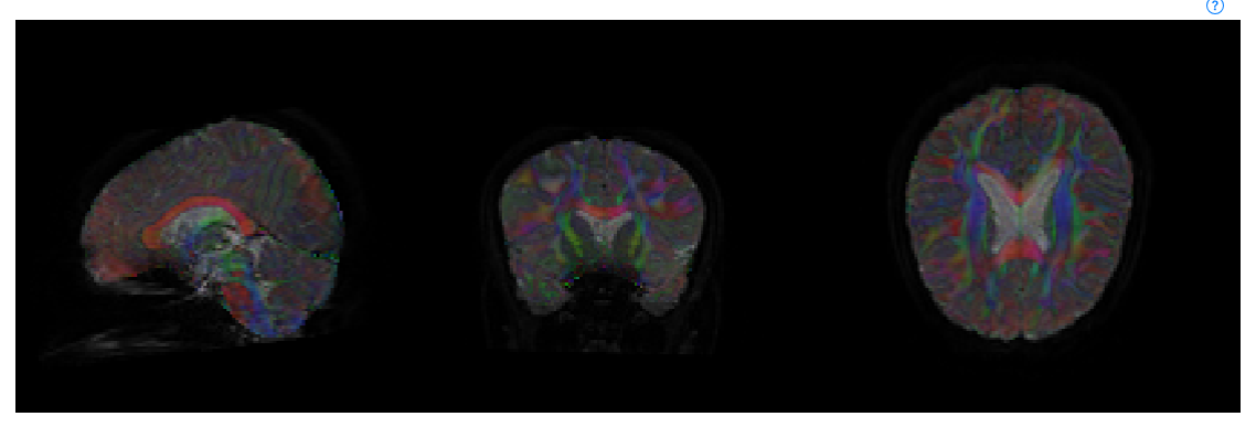
Part 2: Some subjects have color loss/missing signal in the corpus callosum region. Does anyone have ideas for how this came to be, or how we might remedy this?
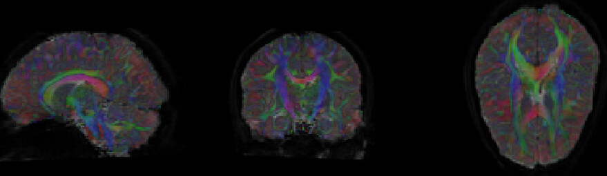
Part 3: We are also noticing a split-color corpus callosum (red/green split where it is typically only red) in some of our scans. We were under the impression that the corpus callosum should typically be a bright red given the directionality of those fibers. However, here you can see half of it is red and half is green (and, in other scans, yellow or pink). We are unsure whether this is a processing error that can be fixed, a result of something more uncontrollable (motion) or an accurate representation of this person’s brain.
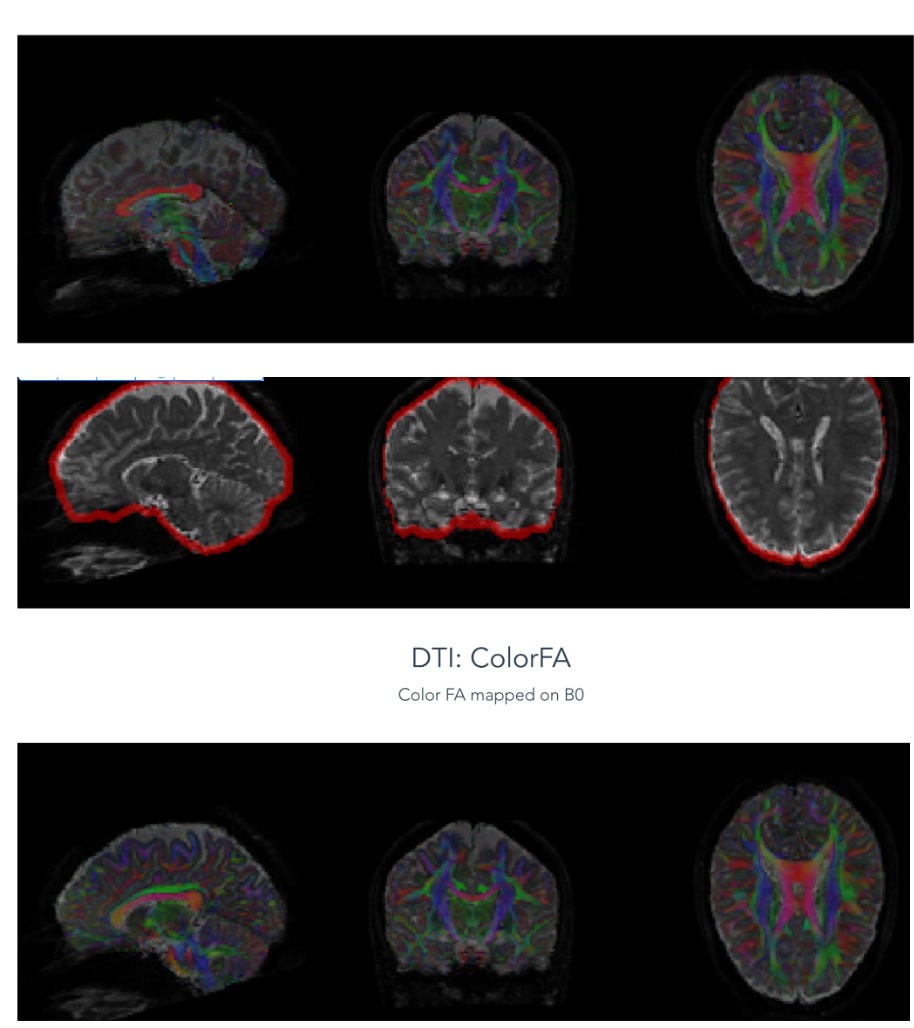
Part 4: In some scans, we see some thick bright whiteness on the top, which seems to coincide with signal loss shown in the motion RMS plot, but could also reflect the subject’s anatomy. Does anyone have an alternate explanation or remedy for this?
Part 5: Lastly, our earlier imaging subjects were run with an 8 channel headcoil, while later subjects were run with a 32 channel headcoil. We have noticed that, across the board, our 8 channel headcoil scans look worse than our 32 channel headcoil scans. The main issue is that, for the earlier scans, we used an older fieldmap workflow where fieldmaps were reconstructed manually from two-echo gradient echo data, converted offline, then passed into QSIPrep, and there may be an issue with how those fieldmaps are scaled or interpreted (or how compatible they are with QSIPrep). Meanwhile, the newer data uses reverse phase-encoding images with TOPUP, a more modern approach for susceptibility distortion correction. Our fix, as of now, is to use fieldmapless distortion correction for the 8 channel headcoil data, which does seem to help a lot. Broadly, is there anything else we should keep in mind when using the 8 channel headcoil data in the future, given the pipeline is now slightly different than the 32 channel headcoil data.
It would help to know more about how you ran QSIPrep, i.e., what is asked for in the Software Support post template. And what kind of acquisition they were.
Yes, that is likely a motion artifact. If you got big signal dropout (e.g., from motion) during a left/right gradient direction, then that would be modeled as artifactual high diffusion during those directions, even if it is not anatomically accurate.
These images are weighted by FA, so that means there is low FA in that region. Not much you can do about that if the quality of the image is poor, unfortunately.
Perhaps the gradient directions are somehow encoded incorrectly in your data? Have you tried running dwigradcheck from MRTrix to see if your gradient table doesn’t have any weird flips in it?
Have you looked at their T1? That would help answer whether it’s artifact vs anatomy.
Are you using SynB0-DisCo or the version built in to QSIPrep? SynB0 might be a promising thing that could work on both images you acquired (if consistency across them is something you need).
It is to say without knowing what you plan on doing with the data, and therefore what kind of biases may or may not be important for you.