Would looking at any of the bval, bvec, and/or json files help? To figure out if there is any other atypicality like the TotalReadoutTimes not being the same?
Thank you so much for your help!!
Would looking at any of the bval, bvec, and/or json files help? To figure out if there is any other atypicality like the TotalReadoutTimes not being the same?
Thank you so much for your help!!
I found a brain that I could share images of here! Images were collected using the same exam card and same sequences as the initial brain I ran and posted about. I ran this brain-to-share through QSIprep using the same command:
qsiprep-docker \Users\harrioem\Desktop\EMHbraindataset \Users\harrioem\Desktop\EMHbraindataset\derivatives -w \Users\harrioem\Desktop\testworkdir --output-resolution 1.3 --fs-license-file \Users\harrioem\Applications\freesurfer\720 --recon-spec pyafq_tractometry
The outputs still look bad. Here is a box link to raw and processed data: Box
I had been looking at these output files: sub-03.html and sub-03_dwiqc.json
Hoping this might help!!
Thank you so very much!
Thanks for sharing the data! I’m not able to download it from the web interface, is it possible to change the permissions?
So sorry! Try again?
Thank you!
Emily
Seems to work now, thanks!
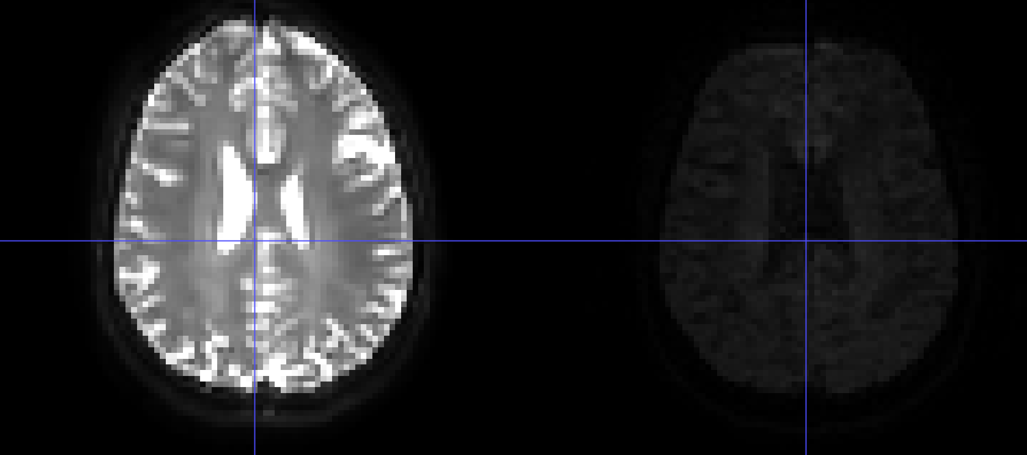
It looks like the image in your fmap directory is not a b=0 image.
This will definitely cause topup to have bad results.
Huh… could the epi.nii.gz have a b=0 image anywhere? Like at the end instead of the usual beginning?
I see that the last volume of epi.nii.gz file has a much higher base magnitude then the other images, so maybe that is the b0?
Yep, I think you’re right! The contrast still looks a little off compared to the b=0 from the dwi
Huh, interesting. So it seems as though my “RPE” image is atypical in its b0 volume placement and perhaps the contrast looks a little weird too. Is there a way I could still use this as an RPE image? By changing the QSIprep inputs/json files or by processing the RPE images first and then using them in QSIprep?
You can try extracting just the last volume of the RPE image and using that as your fieldmap, e.g., with fslsplit and then renaming the last output to the BIDS fieldmap name.
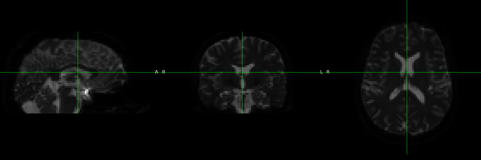
I did: fslsplit sub-03_dir-AP_epi.nii and got 4 output files: vol0000.nii.gz, vol0001.nii.gz, vol0002.nii.gz, vol0003.nii.gz – Would this last one (vol0003.nii.gz) be the “last volume of the RPE image” and should be renamed to the BIDS fieldmap name and used in QSIprep? It also looks different from the other three images (I think more b0-y, closer to the brain on the left side of the screenshot that Matt posted earlier today). See below for a screenshot of vol0003.nii.gz.
Or, alternatively, should I be using fslsplit in fslr instead (R: Split images using FSL)?
Thank you so much!
Yup that is the one!
Okay great!
I deleted my old RPE image from my BIDS folder, copied vol0003.nii.gz into my BIDS folder, renamed vol0003.nii.gz to be sub-03_dir-AP_epi.nii.gz, and ran the same QSIprep command again. BIDS validation passed, and things are running.
I will report back when it’s done. Thank you so much!
Okay! It finished!
IMPORTANT:cli:QSIPrep finished without errors
Without errors, so that’s good. (again, same command as before)
I think the outputs look…better? but still in some places weird? The ventricles look more normally sized, and the colors of the FA map look brighter and more robust and normal, but I think things might still look a little weird (again, looking at sub-03.html and sub-03_dwiqc.json) like the first image (where you look for motion) and the carpet plot and maybe the q space sampling? I uploaded the outputs to a folder on box (use the same link as above: Box) called “18Jan2024NEWFMAP”.
Do you think the outputs also look a little weird? If yes, do you have any recommendations as to what I should try next to make them look more normal? If yes, what should I try next?
Thank you again truly so very much! I am extremely grateful for your help!! ![]()
Have you tried the syn fieldmapless method?
This? Not yet. Is that my most logical next step? Happy to give it a try!
Okay this is what I will try next:
qsiprep-docker \Users\harrioem\Desktop\EMHbraindataset \Users\harrioem\Desktop\EMHbraindataset\derivatives -w \Users\harrioem\Desktop\testworkdir --output-resolution 1.3 --fs-license-file \Users\harrioem\Applications\freesurfer\720 --recon-spec pyafq_tractometry --use-syn-sdc --force-syn --ignore fieldmaps
Will report back when it’s done.
Thank you so much!
Hi @emilymharriott ,
I looked at the dataset and in “18Jan2024NEWFMAP”: both the fmap/*_epi.json file and the dwi/*dwi.json file have "PhaseEncodingDirection": "j". This will not help for distorsion correction. You would want to have for instance "PhaseEncodingDirection": "j-" for sub-03_dir-AP_epi.json.
I hope this helps.
Thank you so very much for your help! Prior to posting here and at the very beginning of this thread, I tried making one j and one j, making one j and one j-, and then making one j- and the other j. Unfortunately, regardless of the - on the j, all the outputs looked the same and also looked bad. Was it still important to have the - on one j?
If so, I’m still running the –use-syn-sdc --force-syn --ignore fieldmaps version (so that shouldn’t matter since I am ignoring fieldmaps) but once I’m done I can try the edited fmap version with j and j-.
MANAGED BY INCF