I am working stroke data and have run my fmriprep pipeline which includes a lesion mask. A lingering question I have prior to running my GLM is whether CompCor produces reliable estimates for stroke data? I just read the Behzadi et al (2007) compcor paper, and after reading have an understanding of what compcor is trying to measure - noise in white matter and CSF. Given that that it should, in theory, help control for CSF related noise in the lesion.
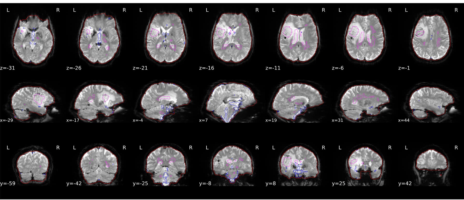
All this being said, I am looking for some guidance on how to look at the figure provided from compcor/fmriprep to decide whether I should actually be using the anatomical CompCor ROI (magenta contour).
Any thoughts, or directions to look for answers would be appreciated!
I wonder if that would work well into excluding the lesion area from segmentation and aCompCor rois calculation.
In general, I would agree with you that signal extracted from the lesion area would have no neuronal components and should be used as confounds for nuisance regression.
Thanks for the reply, did include the lesion mask! So perhaps what I am seeing is CSF in the lesion, which is what the magenta color outlines. Though a slight concern would be that as slices move away from the lesion that tissue that is bordering the lesion that is activating, may be confused with noise.
Usually when questions like this come up, its best to run out the analysis with and without the parameters of question to check for meaningful differences. I’ll do that
When you look at the segmentation masks, is the lesion entirely classified as CSF? From what I understand from the aComCor ROIs calculation, there should not be partial volume effect in the ROI of interest for aCompCor. From the fmriprep boilerplate:
The implementation differs from that of Behzadi et al. in that instead of eroding the masks by 2 pixels on BOLD space, a mask of pixels that likely contain a volume fraction of GM is subtracted from the aCompCor masks. This mask is obtained by dilating a GM mask extracted from the FreeSurfer’s aseg segmentation, and it ensures components are not extracted from voxels containing a minimal fraction of GM. Finally, these masks are resampled into BOLD space and binarized by thresholding at 0.99 (as in the original implementation).
The partial overlap is definitely what had me concerned about using it! The segmentation looks pretty good! There is a bit of lateral space cut off (e.g., z = 19) and a small artifact of lesion circled inside the lesion (see z = 22 and z = 19)
Came across this thread just now while searching for other details about the FMRIPREP aCompCor workflow.
A few years ago I hacked together a script to re-run the aCompCor workflow after excluding voxels within the lesion mask provided to FMRIPREP.
It would likely require some effort to get working with current versions of FMRIPREP, but I figured I’d throw it up on GitHub and post the link here for posterity.