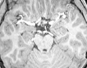
While looking through visual reports generated by MRIQC, I found that many subjects’ T1w images had these white fillings in areas that should be empty. These are especially pronounced in images with low WM2MAX.
What are these white spots? Are they artifacts? If so, what causes them? Will they affect my results (coregistration, functional connectivity)?
A slice from a subject T1w with particularly low WM2MAX
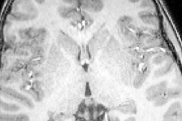
Another slice, same subject. The white fillings are seen throughout most of ventral slices.
The arteries are bright due to flow related enhancement. Different sequences as well as field strength (e.g. while T2 decreases with field strength, T1 increases) can impact this signal. One sequence’s artifacts are another’s signal - a time of flight scan can leverage these flow effects for angiography (with longer T1’s at higher field strength creating better than linear SNR increases with field strength).
I’m really glad you mentioned WM2MAX picks up on these bright arteries, it was actually included into MRIQC with that objective.
Very bright arteries can confuse post-processing steps, specially brain extraction.
What does this mean? It totally depends on the tool and the runtime cost of the methodology. If you run FSL FAST you probably will see some arteries classified as white-matter but if you run FreeSurfer, it’ll probably leave them out. The former takes a few minutes to run and the latter at least 6h for high-quality results.
Oh, I meant fMRIPrep pipeline in particular. Since fMRIPrep uses FSL FAST, it looks like I should watch out for the bright arteries.
Unfortunately, bright arteries are present in quite many images from my dataset, just with different degrees of brightness, so it’s hard to decide where to draw the line for exclusion. I will try processing without exclusion and check the results for low WM2MAX images.