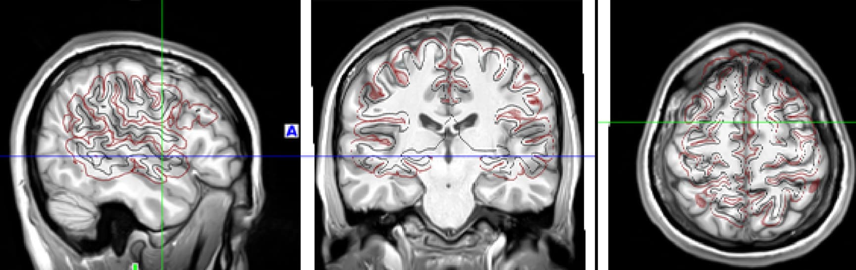
I’m using XCP-D on fMRIprep output. Looking at the executive summary of my subjects, I see that the first images from “BrainSprite Viewer: T1” show misaligned surfaces with respect to the anatomical data:
Data formatted according to a validatable standard? Please provide the output of the validator:
Data is in BIDS standard, but sue to possible misformatted directories, I decided to skip BIDS validation.
Relevant log outputs (up to 20 lines):
N/A
Screenshots / relevant information:
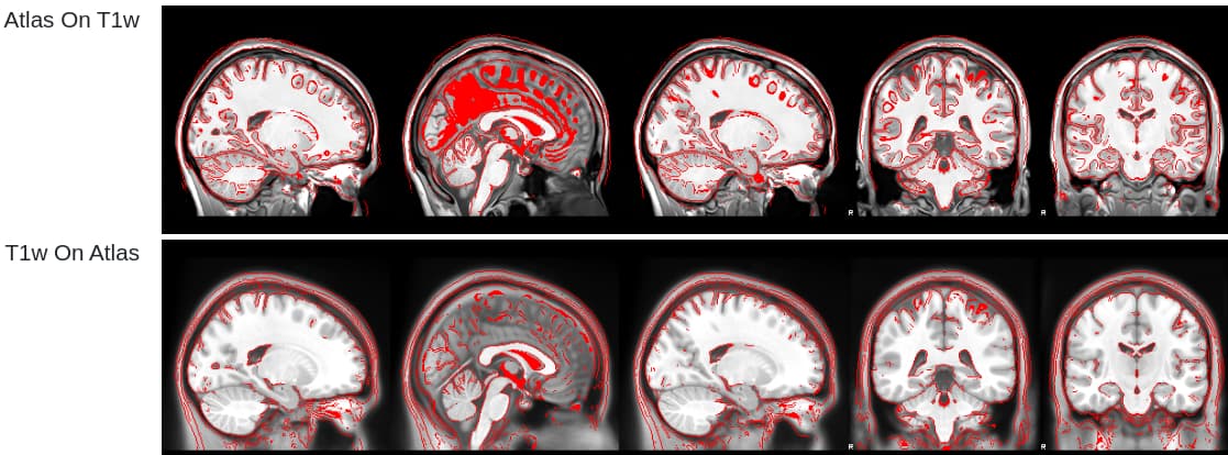
When I load the subject’s FreeSurfer output in freeview, the pial and white-matter surfaces look perfectly fine, suggesting that it’s not an issue of FreeSurfer’s surfaces:
Note that I’m using an older version of XCP-D since I followed this example for using custom confounds. Newer versions of XCP-D use a different syntax for that.
Of course. I repeated the analysis with XCP-D version 0.10.6 but the result stayed the same. The surfaces on top of the T1w images are still misaligned.
I’m not sure what the problem could be. We did see this error in an older version of XCP-D, but that bug should be fixed in the current version. Are you able to share your fMRIPrep derivatives (or perhaps just the raw data from this run)?
Thanks, @tsalo. Certainly. It might be easier if I share with you a minimal portion of the raw data from this run. Perhaps sharing it privately with you by Drive is easiest?
In the meantime, I am rerunning fMRIprep and XCP-D from scratch to make sure no derivatives remain from previous attempts.
While I thought I was running XCP-D 0.10.6, I in fact ran version 0.7.0.
I have now ran version 0.11.1 successfully.
In this version it seems that the summary report does not include any visualization of the anatomical surfaces, so I wasn’t able to verify that the original issue had been resolved. Still, all other outputs seem fine.
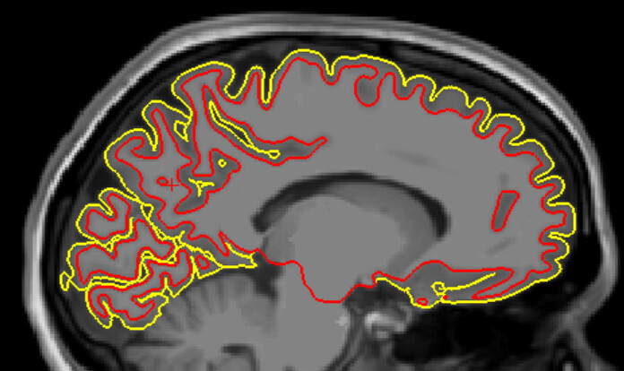
I can confirm that after @tsalo has made some changes in the unstable version 0.11.1, the surfaces are now perfectly aligned. Many thanks for solving this bug, @tsalo!