Summary of what happened:
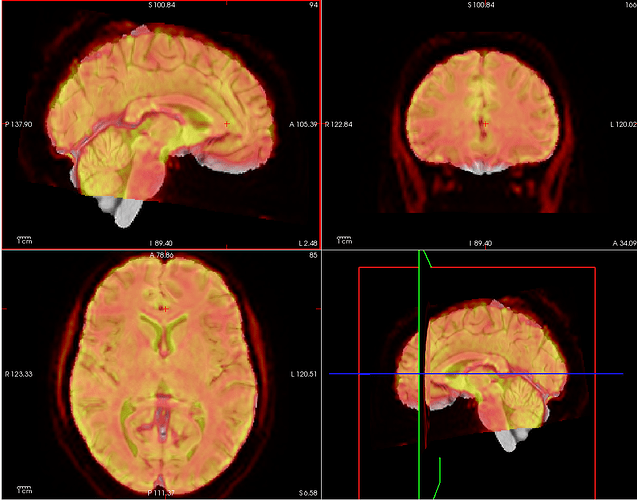
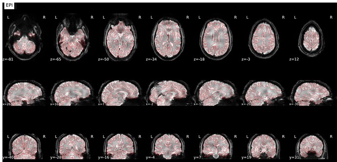
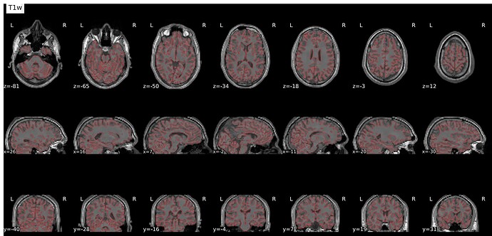
I see a consistent misalignment between the anatomical and functional images generated with fmriprep. (new to fmriprep). I updated the pipeline with some commands I added, but made no difference to the output.
Command used
#!/bin/bash
# User inputs:
bids_root_dir=$HOME/Documents/LUCA/DATA
nthreads=4
mem=25 # in GB
subj=sub-02

# Begin:
echo "Starting the script..."
chmod -R +r /home/maria/Documents/LUCA/DATA/sub-*/func/

# Convert virtual memory from GB to MB
mem=$(echo "${mem//[!0-9]/}") # Remove 'gb' at end
mem_mb=$((mem * 1000 - 5000)) # Reduce some memory for buffer space during pre-processing
echo "Memory allocated: ${mem_mb} MB"

# Set environment variables
export TEMPLATEFLOW_HOME=/home/maria/Documents/LUCA/PREPvalidation/templateflow
export FS_LICENSE=/home/maria/Documents/LUCA/DATA/license.txt
echo "FS License file path: $FS_LICENSE"

# Check if the Docker daemon is running
if ! docker info > /dev/null 2>&1; then
    echo "Docker daemon is not running. Please start Docker."
    exit 1
else
    echo "Docker daemon is running."
fi

echo "Running with Docker..."
docker run --rm -it \
   -v /home/maria/Documents/LUCA/DATA:/Luca \
   -v /media/maria/Storage-8TB/fmriprep_derivatives:/out \
   -v /media/maria/Storage-8TB/BIDSData:/work \
   -v /home/maria/Documents/LUCA/DATA/license.txt:/license.txt:ro \
   nipreps/fmriprep:24.1.1 \
   /Luca /out \
   participant \
   --bold2t1w-dof 9 \
   --bold2t1w-init register \
   --participant-label $subj \
   --skip-bids-validation \
   --fs-license-file /license.txt \
   --output-spaces MNI152NLin2009cAsym fsaverage \
   --use-aroma true \
   --use-syn-sdc \
   --force-bbr \
   --nthreads $nthreads \
   --stop-on-first-crash \
   --mem_mb $mem_mb \
   -w /work

# Check for errors
if [ $? -ne 0 ]; then
    echo "An error occurred while processing participant $subj. Exiting."
    exit 1
fi

echo "All participants processed."
Version:
fmriprep 24.1.1
Environment (Docker, Singularity / Apptainer, custom installation):
Docker
Data formatted according to a validatable standard? Please provide the output of the validator:
BIDS validator shows no errors
Relevant log outputs (up to 20 lines):
No errors to report.
Screenshots / relevant information:
Overlay was made using freeview, I changed the functional image to a heat map and made it translucent. I couldn’t spot a problem with the html report (which I can’t post as I am a new user) but there are some overlapping issues I don’t know how to solve. I would greatly appreciate any help!


