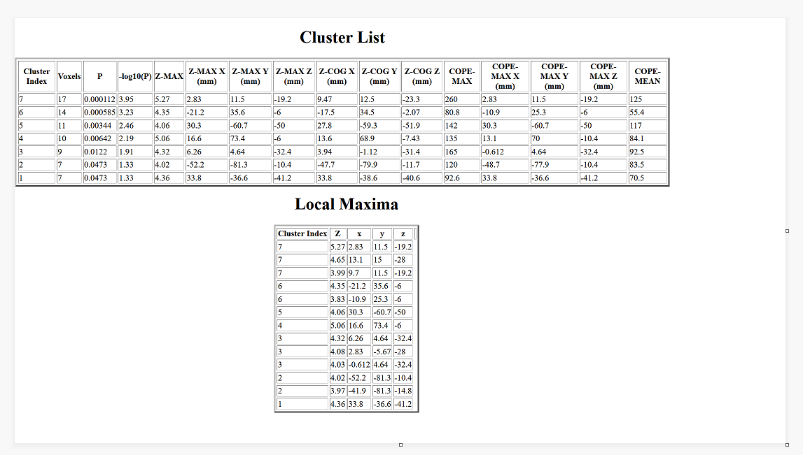
I have a question related to reporting fMRI results using FSL. So, I conducted the first-level and higher-level analysis using FSL and have some results but I don’t know what I should report. Let’s take an example of one contrast. Here’s a screenshot of it.
Wht are you using for results analysis? Is this a single subject or a group? If possible, I would strongly advise using randomize with TFCE. The clsuters look quite small (7 to 17 voxels) and its important to clarify they are cluster level significant. TFCE provides a robust analysis with minimal false positive.
For the table,
p Z cluster size, X, Y, Z, and ideally the BA areas or description of the region.
This result is from a single subject (average of 3 runs).
For the results analysis, I didn’t use anything as yet. This result is from FSL when I click on a single contrast. What do you recommend I use for results analysis? Is there a program/software for TFCE? Where can I get the BA areas?
For the clusters, I would report the Z, X, Y and Z value, and the cope-max of Z-max and would this be for the cluster index with the highest voxels?
I dont wabt to go to far offering advice because i dont use thise fsl tools as much.
TFCE can be implimented in the randonize program. It works at the subject level, but i generally recomment not worrying to much about single subject statistics unless its important for your paper. Its harder to see effecrs at the single subject lwvel, group stats are.more robust.
TFCE is the best correction for multilple comparisons. See Ekund et al, Cluster fail, (sorry on phone cant grab ref).
I think you should report either the cope x y z, or the zmax x y z, not both, they are similar. Just make clear which it is in the table legend.
For BA areas, there are online tools where tou can enter an MNI coordinate andnit guestimates yhe BA region. Also look at online brain atlases and BA overlays in programs like mricron. This is how i leaberned neuroanatomy.