during the acquisition of fMRI Data we, unfortunately, encountered problems with the scanner. To identify resulting artefacts, we used the slice-wise noise average of background data in the fMRI summary plots created by MRI QC.
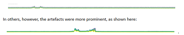
In some participants the artefacts were rather small and not visually detectable in the images, as in the following case:
Are there any indications for acceptable amounts of background noise? Do you have any recommendations on how to proceed?
Since I am working with a clinical population, I also encountered quite a lot of susceptibility artefacts due to metal screws or plates in the head of the patients. Is there an IQM that would be sensitive to these kinds of artefacts, preferably in the functional images?
Lastly, do you know about any reference points for other IQMs (e.g. efc or snr for anatomical and functional images) that would indicate (un-)acceptable values, except for the results of the ABIDE dataset?
I would really appreciate any advice, thank you so much in advance!
Interesting observation thank you for sharing! If you can, could you show us how the artefact manifest themselves on the images (anatomical and/or functional) in the second case (where it is more visible in the slice-wise noise average plot?)
By definition the susceptibility artefact from metals implants would show strong geometric deformation and dropout of signal in more or less close proximity of the metallic part (depending on the size and composition of the metallic part, as well as the type of MRI sequences used and their acquisition parameters) . I am not totally sure which IQM would be most sensitive to this. In your visual reports, in the T1w, is the brainmask affected by the artefacts?
This is a a wild guess: perhaps in presence of of this artefact, the FD curves have a different shape, due to the interaction of the real motion and the susceptibility artefact? Something also maybe visible in the carpet plot? Some voxels wrongly affected to a certain tissue type (interaction of the susceptibility artefact with the segmentation) and this voxels may have a weird temporal evolution?
Otherwise, looking at the IQMs themselves, you will get some outliers in the T1w reports (summary_bg) and perhaps in the functional ones (aor, aqi).
In general, I would recommend to start by looking at your group reports and look for outliers in any metrics and check if you can find a group of outliers that corresponds to the images of participants with metallic artefacts.
Hi,
thank you so much for your reply!
The scanner artefacts only show in the DICOM files. It’s a little hard to see on just one picture, but you can see a white line on the right side of the image. This white line basically just moved through the whole image over time in the functional scans. I followed your advice and looked at the group plots to identify in which IQM the affected subjecs scored differently than the others. The summary_fg reports of the bold images seemed to be quite sensitive.