Hi everyone,
I’m a beginner in fMRI analysis, and our team is currently facing an issue related to data acquisition and preprocessing.
While reviewing our fMRI task data, we noticed that some of the functional scans were stored in sagittal view, while others including both functional scans and T1-weighted images were stored in axial view. It appears that the MRI protocol settings were changed at some point, although we’re not exactly sure why.
I’m wondering whether this inconsistency in storage orientation could lead to any problems during preprocessing or subsequent analysis. For example, if I understand correctly, this might affect the coregistration step, since our T1-weighted images were acquired in axial view, whereas some of our EPI data were acquired in sagittal view.
Would it still be possible to use the sagittal view data, or would it be better to exclude them? If there’s a recommended way to address this, I’d greatly appreciate your advice.
Mijeong
Howdy-
Just to check, are the data appearing to be reoriented improperly in space? Or is the header information for the datasets showing that the dataset orientation is different?
That is, do all the data display correctly in a GUI (I would use the AFNI GUI, for example), including overlap? And is the only difference observable with header checks, like:
3dinfo -orient -prefix DSET1 [DSET2 DSET3 ...]
?
Note that there is a program in AFNI called gtkyd_check.py, where “gtkyd” stands for “Getting to Know Your Data”. This is useful for agglomerating a lot of header info across a batch of data into a CSV or spreadsheet file, and then you can followup and check it for properties (even programmatically with gen_ss_review_table.py—see gtkyd_check.py’s examples in the help file). This would help you assemble all the header info for all your EPI data, and separately for your anatomical data.
Also, are you sure from looking at your raw dicoms that the datasets were really acquired in different slice places? For EPI data, that miiiight affect distortions, creating a difference across subjects. For anatomical data, without any other information I wouldn’t expect it to cause an issue. However, sometimes programs that convert DICOM->NIFTI might reorient the data just on the disk, meaning that the actual data is not different, and something like 3dresample could be used to correct/make uniform the dataset orientations. But it depends on where the orientation difference is actually occurring—in the original acquisition, or in the DICOM->NIFTI conversion step.
–pt
I think Ben Inglis does a nice job of discussing EPI artifacts for axial as well as coronal, and sagittal scans. In general, each direction has its own tradeoffs, but mixing different directions in a single study may impact statistical power by adding a lot of variance. A good method for reducing geometric distortions (e.g. a field map or reversed phase encoding sequence) might minimize these artifacts and lead to more homogenous data.
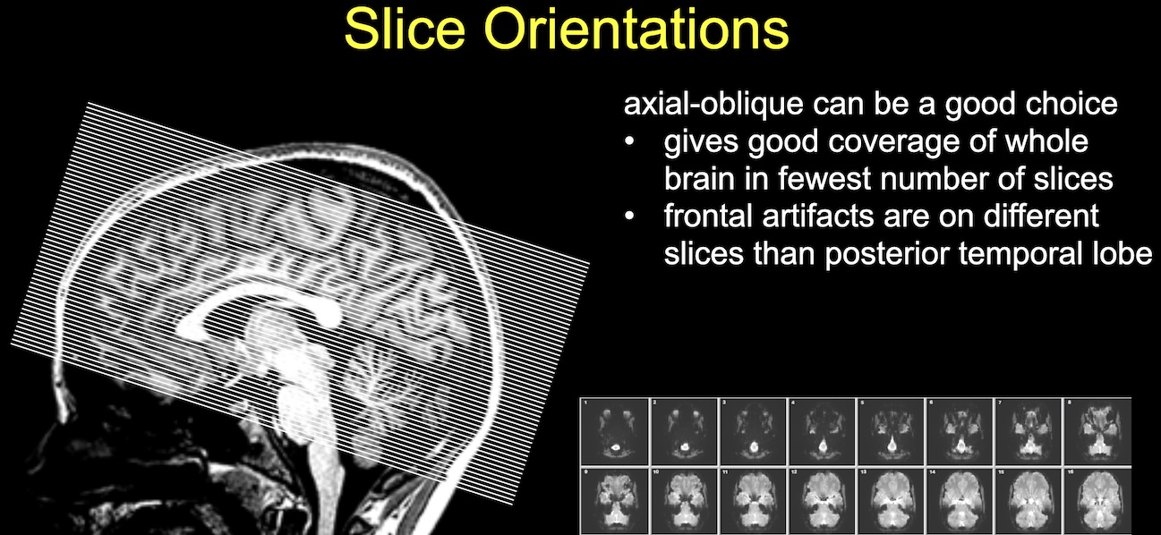
As Jody Culham notes in Lecture 11c: fMRI Physics, an axial-oblique slice direction also has its benefits: