Hi, I am trying to adopt new 7t machine for fMRI analysis, and having trouble with spatial normalization using fmriprep.
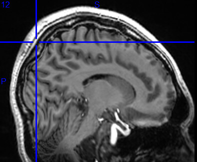
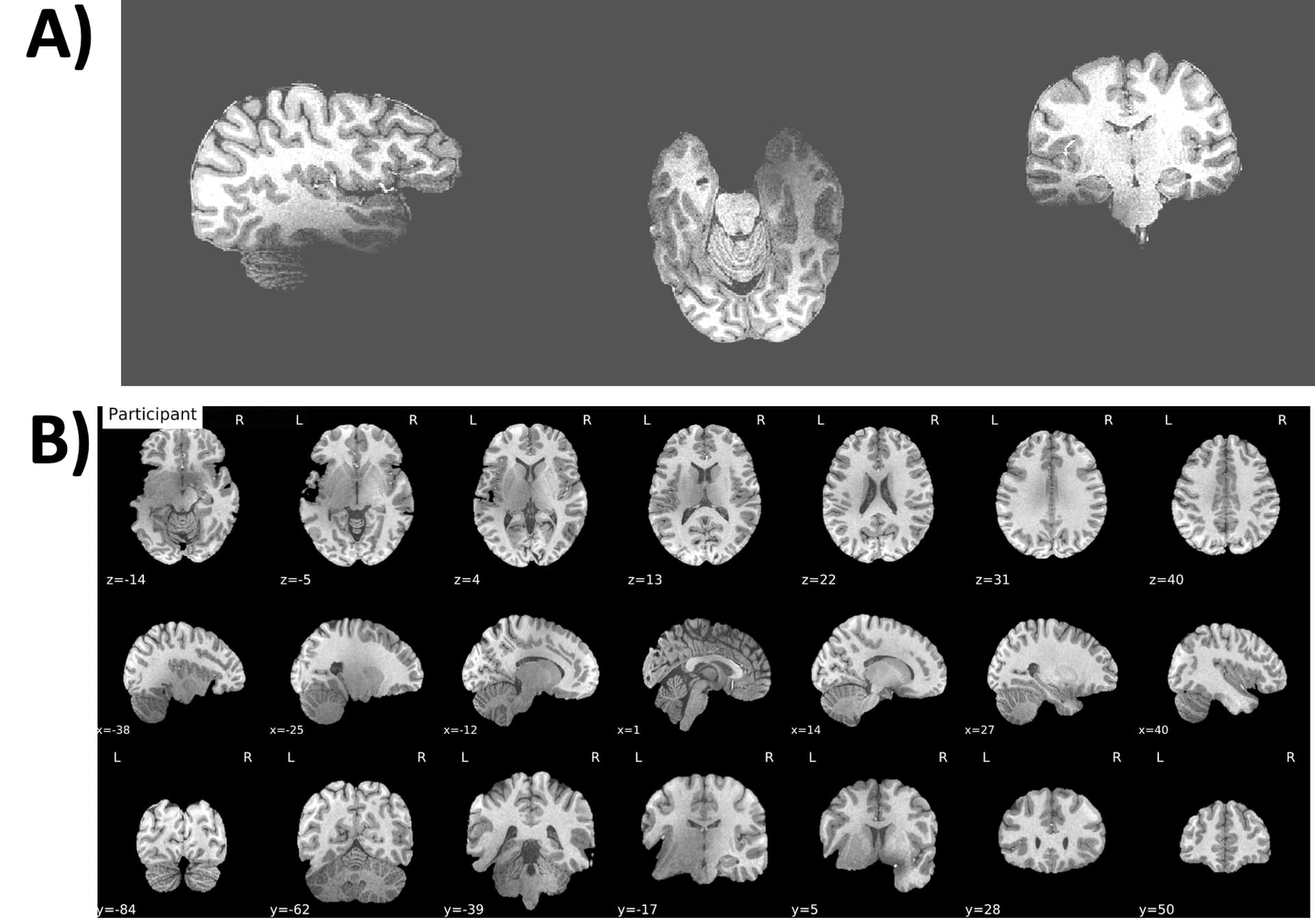
As you can see from attached data, occipital area and cerebellum region are visibly distorted.
This issue does not show when we use 3t anatomical image for analysis, so it seems like a issue might be the 7t anatomical image scans…
How can I fix the problem with the data? Or is there anything I need to check or fix when we scanning the participants to solve this issue? Any answers would be really helpful. Thanks!
Hi @leeeut000, I assume that the image looks reasonable prior to using with fMRIPrep?
How do the other steps perform? Often bad normalization results from a bad brain mask. We may also need to update the registration parameters.
Bringing fMRIPrep up to date to work with 7T images might be a non-trivial effort. Would you be interested in being involved? We’d be happy to have your contributions and help you getting started with development.
We have noticed that the intensity of our anatomical images really drops off in the temporal lobes, exactly where most of the distortions are happening. This is due to field inhomogeneities present at 7T. Is it possible this is related to an intensity normalization step during recon-all?