Hi,
Ghosting is inherent to echo planar imaging (EPI) acquisition. Visually, just from this image it is difficult to tell if it is worse than usual, but it seems normal to me.
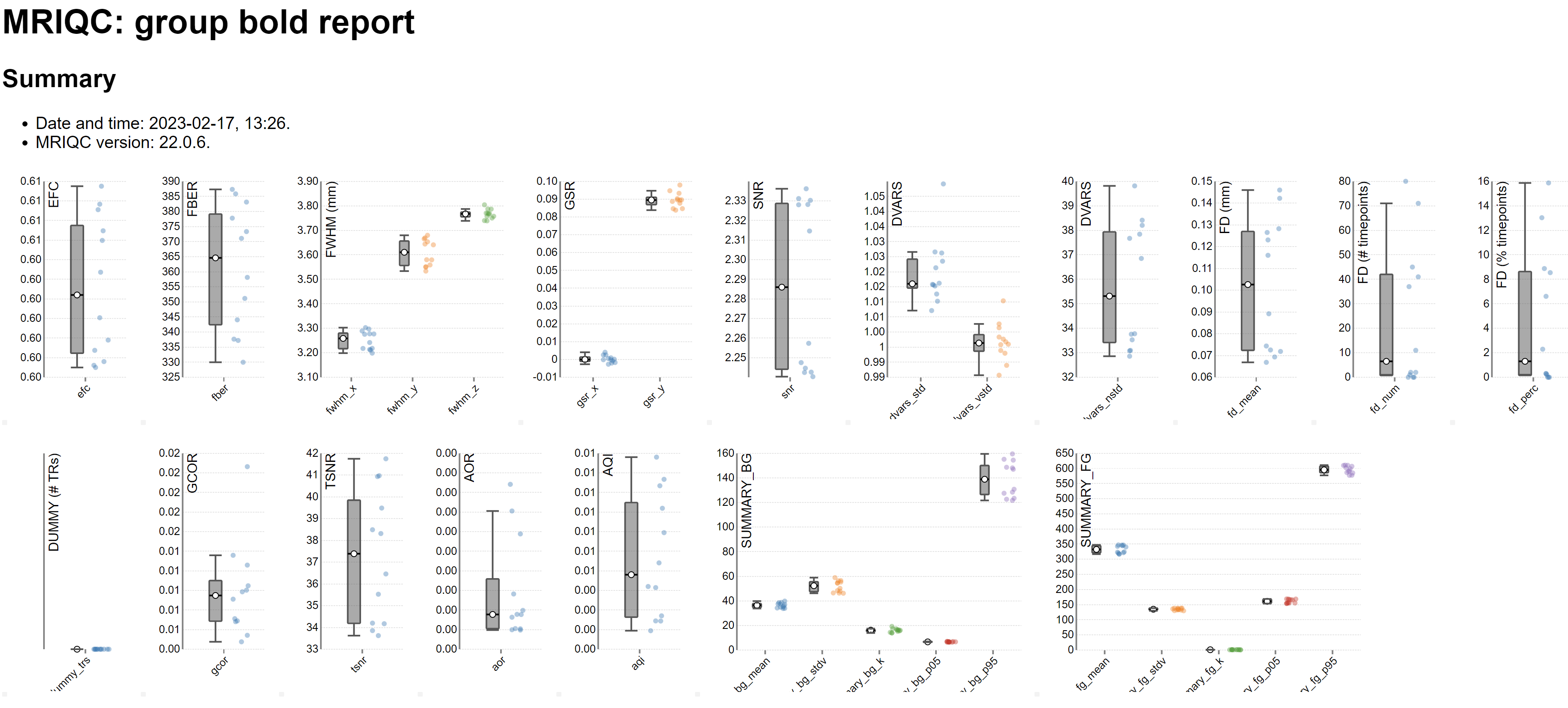
It is possible to assess this artefact quantitatively with metrics such as Ghost to Signal Ratio implemented in tools like MRIQC:
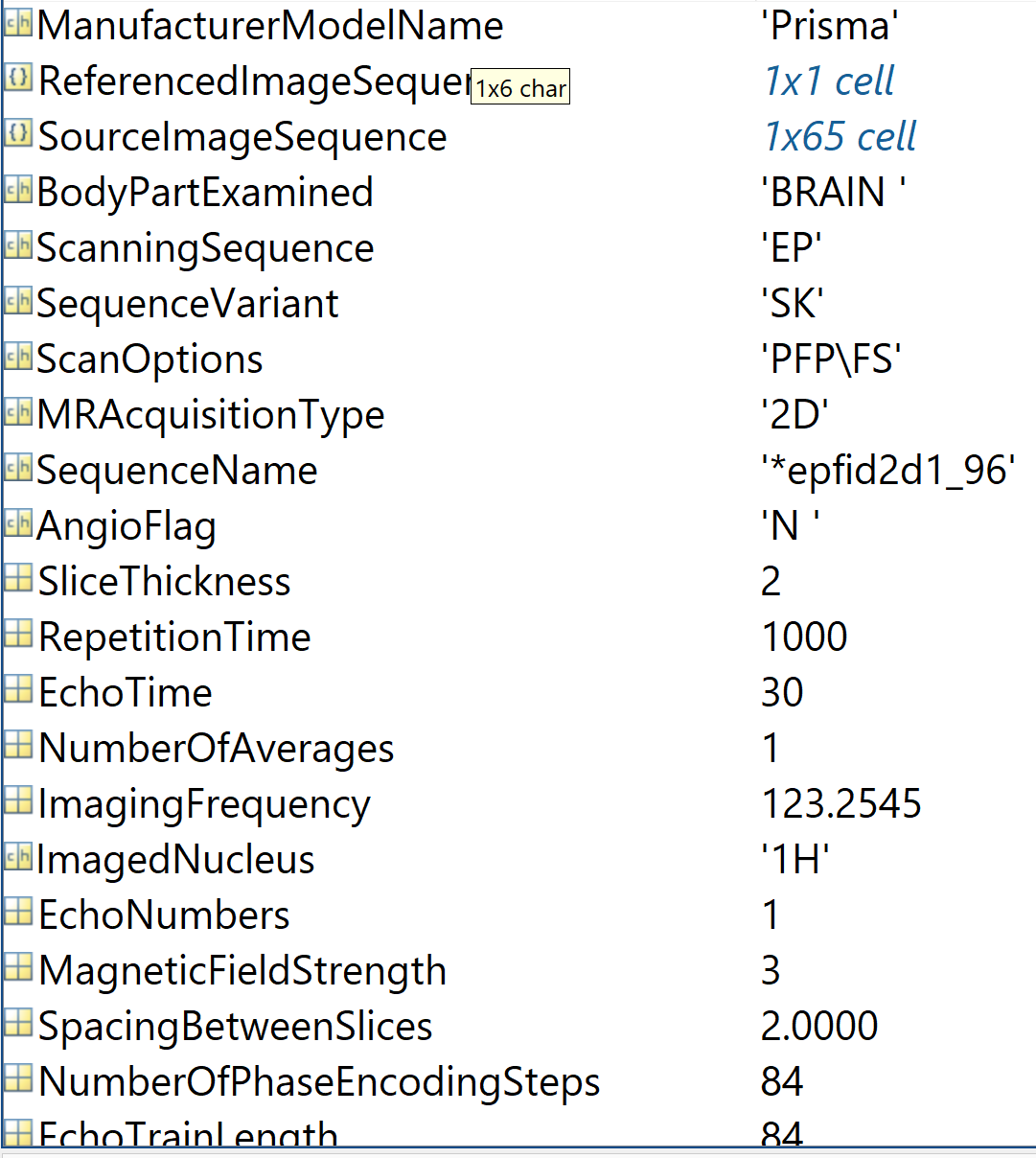
how many slices did you use? It looks like you have a TR of 1s, with slices of 2mm. Do you use any kind of acceleration? (in-plane (GRAPPA) or multi-slice?)
You use a flip angle of 90 degrees which is not optimal for such a short TR, the Ernst Angle would be about 62 degrees. (taking a T1 of GM of 1300ms). Some studies recommend to lower even more the flip angle to reduce the physiological noise (Gonzalez-Castillo et al. 2011)
We used 65 slices and yes, we used multi-slice acceleration.
Thanks for your suggestion. Should I consider other parameters if fine-tuning to smaller flip angle? Sorry that I know very little about the physical principles of MRI.
Judging how much ghosting is “too much” is not straightforward, and (as usual) depends on multiple considerations, including the type of analysis you’re planning. Definitely ask your colleagues, imaging center staff, physicists, etc. for their opinion of your pilot images (and acquisition parameters). Looking at other datasets can also help to train your eye, e.g., OSF.
If at all possible, include some control analyses (tasks known to produce strong, consistent responses in the brain areas of most interest to you) in your piloting, so you can confirm that the acquisition produces reasonably clear signal.